[English] 日本語
Yorodumi
- EMDB-30007: Cryo-EM structure of phosphoketolase from Bifidobacterium longum -

+
Open data


ID or keywords:

Loading...

-
Basic information

Entry
Database: EMDB / ID: EMD-30007
TitleCryo-EM structure of phosphoketolase from Bifidobacterium longum
Map data
Sample
  • Complex: Phosphoketolase with thiamine-diphophate
    • Protein or peptide: Phosphoketolase
  • Ligand: THIAMINE DIPHOSPHATEThiamine pyrophosphate
  • Ligand: CALCIUM IONCalcium
  • Ligand: water
Function / homology
Function and homology information


fructose-6-phosphate phosphoketolase / fructose-6-phosphate phosphoketolase activity / carbohydrate metabolic process
Similarity search - Function
Xylulose 5-phosphate/Fructose 6-phosphate phosphoketolase / Xylulose 5-phosphate/Fructose 6-phosphate phosphoketolase, C-terminal / Xylulose 5-phosphate/Fructose 6-phosphate phosphoketolase, N-terminal / Xylulose 5-phosphate/Fructose 6-phosphate phosphoketolase, thiamine diphosphate binding site / Xylulose 5-phosphate/Fructose 6-phosphate phosphoketolase, conserved site / D-xylulose 5-phosphate/D-fructose 6-phosphate phosphoketolase / XFP C-terminal domain / XFP N-terminal domain / Phosphoketolase signature 1. / Phosphoketolase signature 2. ...Xylulose 5-phosphate/Fructose 6-phosphate phosphoketolase / Xylulose 5-phosphate/Fructose 6-phosphate phosphoketolase, C-terminal / Xylulose 5-phosphate/Fructose 6-phosphate phosphoketolase, N-terminal / Xylulose 5-phosphate/Fructose 6-phosphate phosphoketolase, thiamine diphosphate binding site / Xylulose 5-phosphate/Fructose 6-phosphate phosphoketolase, conserved site / D-xylulose 5-phosphate/D-fructose 6-phosphate phosphoketolase / XFP C-terminal domain / XFP N-terminal domain / Phosphoketolase signature 1. / Phosphoketolase signature 2. / Transketolase C-terminal/Pyruvate-ferredoxin oxidoreductase domain II / Thiamin diphosphate-binding fold
Similarity search - Domain/homology
Biological speciesBifidobacterium longum subsp. longum F8 (bacteria)
Methodsingle particle reconstruction / cryo EM / Resolution: 2.1 Å
AuthorsNakata K / Miyazaki N / Yamaguchi H / Hirose M / Miyano H / Mizukoshi T / Kashiwagi T / Iwasaki K
Funding support Japan, 1 items
OrganizationGrant numberCountry
Japan Agency for Medical Research and Development (AMED) Japan
Citation
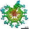
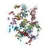
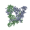
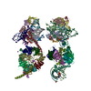
Journal: J Struct Biol / Year: 2022
Title: High-resolution structure of phosphoketolase from Bifidobacterium longum determined by cryo-EM single-particle analysis.
Authors: Kunio Nakata / Naoyuki Miyazaki / Hiroki Yamaguchi / Mika Hirose / Tatsuki Kashiwagi / Nidamarthi H V Kutumbarao / Osamu Miyashita / Florence Tama / Hiroshi Miyano / Toshimi Mizukoshi / Kenji Iwasaki /
Abstract: In bifidobacteria, phosphoketolase (PKT) plays a key role in the central hexose fermentation pathway called "bifid shunt." The three-dimensional structure of PKT from Bifidobacterium longum with co- ...In bifidobacteria, phosphoketolase (PKT) plays a key role in the central hexose fermentation pathway called "bifid shunt." The three-dimensional structure of PKT from Bifidobacterium longum with co-enzyme thiamine diphosphate (ThDpp) was determined at 2.1 Å resolution by cryo-EM single-particle analysis using 196,147 particles to build up the structural model of a PKT octamer related by D symmetry. Although the cryo-EM structure of PKT was almost identical to the X-ray crystal structure previously determined at 2.2 Å resolution, several interesting structural features were observed in the cryo-EM structure. Because this structure was solved at relatively high resolution, it was observed that several amino acid residues adopt multiple conformations. Among them, Q546-D547-H548-N549 (the QN-loop) demonstrate the largest structural change, which seems to be related to the enzymatic function of PKT. The QN-loop is at the entrance to the substrate binding pocket. The minor conformer of the QN-loop is similar to the conformation of the QN-loop in the crystal structure. The major conformer is located further from ThDpp than the minor conformer. Interestingly, the major conformer in the cryo-EM structure of PKT resembles the corresponding loop structure of substrate-bound Escherichia coli transketolase. That is, the minor and major conformers may correspond to "closed" and "open" states for substrate access, respectively. Moreover, because of the high-resolution analysis, many water molecules were observed in the cryo-EM structure of PKT. Structural features of the water molecules in the cryo-EM structure are discussed and compared with water molecules observed in the crystal structure.
History
DepositionFeb 12, 2020-
Header (metadata) releaseFeb 17, 2021-
Map releaseFeb 17, 2021-
UpdateOct 12, 2022-
Current statusOct 12, 2022Processing site: PDBj / Status: Released

-
Structure visualization
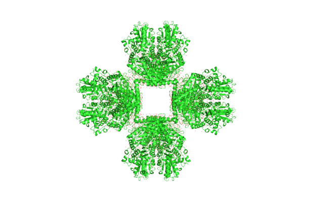
Movie
  • Surface view with section colored by density value
  • Surface level: 0.05
  • Imaged by UCSF Chimera
  • Download
  • Surface view colored by cylindrical radius
  • Surface level: 0.05
  • Imaged by UCSF Chimera
  • Download
  • Surface view with fitted model
  • Atomic models: PDB-6lxv
  • Surface level: 0.05
  • Imaged by UCSF Chimera
  • Download
  • Simplified surface model + fitted atomic model
  • Atomic modelsPDB-6lxv
  • Imaged by Jmol
  • Download
Movie viewer
Structure viewerEM map:
SurfViewMolmilJmol/JSmol
Supplemental images

Downloads & links

-
Map

FileDownload / File: emd_30007.map.gz / Format: CCP4 / Size: 178 MB / Type: IMAGE STORED AS FLOATING POINT NUMBER (4 BYTES)
Voxel sizeX=Y=Z: 0.87 Å
Density
Contour LevelBy AUTHOR: 0.05 / Movie #1: 0.05
Minimum - Maximum-0.20026584 - 0.54527557
Average (Standard dev.)0.00035448186 (±0.012134807)
SymmetrySpace group: 1
Details

EMDB XML:

Map geometry
Axis orderXYZ
Origin000
Dimensions360360360
Spacing360360360
CellA=B=C: 313.2 Å
α=β=γ: 90.0 °

CCP4 map header:

modeImage stored as Reals
Å/pix. X/Y/Z0.870.870.87
M x/y/z360360360
origin x/y/z0.0000.0000.000
length x/y/z313.200313.200313.200
α/β/γ90.00090.00090.000
start NX/NY/NZ000
NX/NY/NZ256256256
MAP C/R/S123
start NC/NR/NS000
NC/NR/NS360360360
D min/max/mean-0.2000.5450.000

-
Supplemental data

-
Sample components

-
Entire : Phosphoketolase with thiamine-diphophate

EntireName: Phosphoketolase with thiamine-diphophate
Components
  • Complex: Phosphoketolase with thiamine-diphophate
    • Protein or peptide: Phosphoketolase
  • Ligand: THIAMINE DIPHOSPHATEThiamine pyrophosphate
  • Ligand: CALCIUM IONCalcium
  • Ligand: water

-
Supramolecule #1: Phosphoketolase with thiamine-diphophate

SupramoleculeName: Phosphoketolase with thiamine-diphophate / type: complex / ID: 1 / Parent: 0 / Macromolecule list: #1
Source (natural)Organism: Bifidobacterium longum subsp. longum F8 (bacteria)
Recombinant expressionOrganism: Escherichia coli BL21(DE3) (bacteria) / Recombinant plasmid: pET24

-
Macromolecule #1: Phosphoketolase

MacromoleculeName: Phosphoketolase / type: protein_or_peptide / ID: 1 / Number of copies: 8 / Enantiomer: LEVO / EC number: fructose-6-phosphate phosphoketolase
Source (natural)Organism: Bifidobacterium longum subsp. longum F8 (bacteria)
Molecular weightTheoretical: 93.450016 KDa
Recombinant expressionOrganism: Escherichia coli BL21(DE3) (bacteria)
SequenceString: MTSPVIGTPW KKLNAPVSEE ALEGVDKYWR VANYLSIGQI YLRSNPLMKE PFTREDVKHR LVGHWGTTPG LNFLIGHINR FIADHGQNT VIIMGPGHGG PAGTSQSYLD GTYTETFPKI TKDEAGLQKF FRQFSYPGGI PSHFAPETPG SIHEGGELGY A LSHAYGAI ...String:
MTSPVIGTPW KKLNAPVSEE ALEGVDKYWR VANYLSIGQI YLRSNPLMKE PFTREDVKHR LVGHWGTTPG LNFLIGHINR FIADHGQNT VIIMGPGHGG PAGTSQSYLD GTYTETFPKI TKDEAGLQKF FRQFSYPGGI PSHFAPETPG SIHEGGELGY A LSHAYGAI MDNPSLFVPA IVGDGEAETG PLATGWQSNK LVNPRTDGIV LPILHLNGYK IANPTILSRI SDEELHEFFH GM GYEPYEF VAGFDDEDHM SIHRRFAELW ETIWDEICDI KATAQTDNVH RPFYPMLIFR TPKGWTCPKY IDGKKTEGSW RSH QVPLAS ARDTEAHFEV LKNWLESYKP EELFDANGAV KDDVLAFMPK GELRIGANPN ANGGVIRNDL KLPNLEDYEV KEVA EYGHG WGQLEATRTL GAYTRDIIKN NPRDFRIFGP DETASNRLQA SYEVTNKQWD AGYISDEVDE HMHVSGQVVE QLSEH QMEG FLEAYLLTGR HGIWSSYESF VHVIDSMLNQ HAKWLEATVR EIPWRKPIAS MNLLVSSHVW RQDHNGFSHQ DPGVTS VLL NKCFHNDHVI GIYFATDANM LLAIAEKCYK STNKINAIIA GKQPAATWLT LDEARAELEK GAAAWDWAST AKNNDEA EV VLAAAGDVPT QEIMAASDKL KELGVKFKVV NVADLLSLQS AKENDEALTD EEFADIFTAD KPVLFAYHSY AHDVRGLI Y DRPNHDNFNV HGYEEEGSTT TPYDMVRVNR IDRYELTAEA LRMIDADKYA DKIDELEKFR DEAFQFAVDN GYDHPDYTD WVYSGVNTDK KGAVTATAAT AGDNEHHHHH H

-
Macromolecule #2: THIAMINE DIPHOSPHATE

MacromoleculeName: THIAMINE DIPHOSPHATE / type: ligand / ID: 2 / Number of copies: 8 / Formula: TPP
Molecular weightTheoretical: 425.314 Da
Chemical component information

ChemComp-TPP:
THIAMINE DIPHOSPHATE / Thiamine pyrophosphate

-
Macromolecule #3: CALCIUM ION

MacromoleculeName: CALCIUM ION / type: ligand / ID: 3 / Number of copies: 8 / Formula: CA
Molecular weightTheoretical: 40.078 Da

-
Macromolecule #4: water

MacromoleculeName: water / type: ligand / ID: 4 / Number of copies: 2967 / Formula: HOH
Molecular weightTheoretical: 18.015 Da
Chemical component information

ChemComp-HOH:
WATER / Water

-
Experimental details

-
Structure determination

Methodcryo EM
Processingsingle particle reconstruction
Aggregation stateparticle

-
Sample preparation

Concentration10 mg/mL
BufferpH: 9
GridModel: Quantifoil R1.2/1.3 / Material: MOLYBDENUM / Mesh: 300 / Pretreatment - Type: GLOW DISCHARGE
VitrificationCryogen name: ETHANE

-
Electron microscopy

MicroscopeFEI TITAN KRIOS
Electron beamAcceleration voltage: 300 kV / Electron source: FIELD EMISSION GUN
Electron opticsIllumination mode: FLOOD BEAM / Imaging mode: BRIGHT FIELDBright-field microscopy / Nominal defocus max: 2.75 µm / Nominal defocus min: 1.0 µm / Nominal magnification: 75000
Sample stageSpecimen holder model: FEI TITAN KRIOS AUTOGRID HOLDER / Cooling holder cryogen: NITROGEN
Image recordingFilm or detector model: FEI FALCON III (4k x 4k) / Detector mode: COUNTING / Digitization - Dimensions - Width: 4096 pixel / Digitization - Dimensions - Height: 4096 pixel / Number real images: 2897 / Average exposure time: 45.0 sec. / Average electron dose: 50.0 e/Å2
Experimental equipment
Model: Titan Krios / Image courtesy: FEI Company

-
Image processing

Particle selectionNumber selected: 426149
CTF correctionSoftware - Name: Gctf (ver. 1.06)
Initial angle assignmentType: OTHER / Software - Name: RELION (ver. 3.0)
Final 3D classificationNumber classes: 4 / Avg.num./class: 50000 / Software - Name: RELION (ver. 3.0)
Final angle assignmentType: MAXIMUM LIKELIHOOD / Software - Name: RELION (ver. 3.0)
Final reconstructionApplied symmetry - Point group: D4 (2x4 fold dihedral) / Algorithm: FOURIER SPACE / Resolution.type: BY AUTHOR / Resolution: 2.1 Å / Resolution method: FSC 0.143 CUT-OFF / Software - Name: RELION (ver. 3.0) / Number images used: 194517
FSC plot (resolution estimation)

+
About Yorodumi

-
News

-
Feb 9, 2022. New format data for meta-information of EMDB entries

New format data for meta-information of EMDB entries

  • Version 3 of the EMDB header file is now the official format.
  • The previous official version 1.9 will be removed from the archive.

Related info.:EMDB header

External links:wwPDB to switch to version 3 of the EMDB data model

-
Aug 12, 2020. Covid-19 info

Covid-19 info

URL: https://pdbj.org/emnavi/covid19.php

New page: Covid-19 featured information page in EM Navigator.

Related info.:Covid-19 info / Mar 5, 2020. Novel coronavirus structure data

+
Mar 5, 2020. Novel coronavirus structure data

Novel coronavirus structure data

Related info.:Yorodumi Speices / Aug 12, 2020. Covid-19 info

External links:COVID-19 featured content - PDBj / Molecule of the Month (242):Coronavirus Proteases

+
Jan 31, 2019. EMDB accession codes are about to change! (news from PDBe EMDB page)

EMDB accession codes are about to change! (news from PDBe EMDB page)

  • The allocation of 4 digits for EMDB accession codes will soon come to an end. Whilst these codes will remain in use, new EMDB accession codes will include an additional digit and will expand incrementally as the available range of codes is exhausted. The current 4-digit format prefixed with “EMD-” (i.e. EMD-XXXX) will advance to a 5-digit format (i.e. EMD-XXXXX), and so on. It is currently estimated that the 4-digit codes will be depleted around Spring 2019, at which point the 5-digit format will come into force.
  • The EM Navigator/Yorodumi systems omit the EMD- prefix.

Related info.:Q: What is EMD? / ID/Accession-code notation in Yorodumi/EM Navigator

External links:EMDB Accession Codes are Changing Soon! / Contact to PDBj

+
Jul 12, 2017. Major update of PDB

Major update of PDB

  • wwPDB released updated PDB data conforming to the new PDBx/mmCIF dictionary.
  • This is a major update changing the version number from 4 to 5, and with Remediation, in which all the entries are updated.
  • In this update, many items about electron microscopy experimental information are reorganized (e.g. em_software).
  • Now, EM Navigator and Yorodumi are based on the updated data.

External links:wwPDB Remediation / Enriched Model Files Conforming to OneDep Data Standards Now Available in the PDB FTP Archive

-
Yorodumi

Thousand views of thousand structures

  • Yorodumi is a browser for structure data from EMDB, PDB, SASBDB, etc.
  • This page is also the successor to EM Navigator detail page, and also detail information page/front-end page for Omokage search.
  • The word "yorodu" (or yorozu) is an old Japanese word meaning "ten thousand". "mi" (miru) is to see.

Related info.:EMDB / PDB / SASBDB / Comparison of 3 databanks / Yorodumi Search / Aug 31, 2016. New EM Navigator & Yorodumi / Yorodumi Papers / Jmol/JSmol / Function and homology information / Changes in new EM Navigator and Yorodumi

Read more