 Movie
Movie Controller

[English] 日本語
 Yorodumi
- PDB-6ofb: Crystal structure of human glutamine-dependent NAD+ synthetase co... -
+ Open data

- Basic information
| Entry | Database: PDB / ID: 6ofb | ||||||
|---|---|---|---|---|---|---|---|
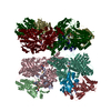
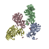
| Title | Crystal structure of human glutamine-dependent NAD+ synthetase complexed with NaAD+, AMP, pyrophosphate, and Mg2+ | ||||||
 Components Components | Glutamine-dependent NAD(+) synthetase | ||||||
 Keywords Keywords |  LIGASE / GLUTAMINE-AMIDO TRANSFERASE / GAT / NAD+ SYNTHETASE / GLUTAMINE-DEPENDENT NAD+ SYNTHETASE / NAD SYNTHETASE 1 / GLUTAMINASE / AMMONIA TUNNELING / ENZYME / ATP-BINDING / NUCLEOTIDE-BINDING LIGASE / GLUTAMINE-AMIDO TRANSFERASE / GAT / NAD+ SYNTHETASE / GLUTAMINE-DEPENDENT NAD+ SYNTHETASE / NAD SYNTHETASE 1 / GLUTAMINASE / AMMONIA TUNNELING / ENZYME / ATP-BINDING / NUCLEOTIDE-BINDING | ||||||
| Function / homology |  Function and homology information Function and homology information'de novo' NAD biosynthetic process / NAD+ synthase (glutamine-hydrolysing) / NAD+ synthase (glutamine-hydrolyzing) activity / Nicotinate metabolism / NAD metabolic process / glutaminase activity / NAD biosynthetic process / ATP binding / cytosol / cytoplasmSimilarity search - Function | ||||||
| Biological species |  Homo sapiens (human) Homo sapiens (human) | ||||||
| Method | X-RAY DIFFRACTION / SYNCHROTRON / MOLECULAR REPLACEMENT / Resolution: 2.84 Å | ||||||
 Authors Authors | Chuenchor, W. / Doukov, T.I. / Gerratana, B. | ||||||
 Citation Citation | Journal: Nat Commun / Year: 2020 Title: Different ways to transport ammonia in human and Mycobacterium tuberculosis NAD+synthetases. Authors: Chuenchor, W. / Doukov, T.I. / Chang, K.T. / Resto, M. / Yun, C.S. / Gerratana, B. | ||||||
| History |
|
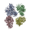
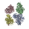
- Structure visualization
| Structure viewer | Molecule: MolmilJmol/JSmol |
|---|
- Downloads & links
-Download
| PDBx/mmCIF format | 6ofb.cif.gz | 354.6 KB | Display | PDBx/mmCIF format |
|---|---|---|---|---|
| PDB format | pdb6ofb.ent.gz | 230.9 KB | Display | PDB format |
| PDBx/mmJSON format | 6ofb.json.gz | Tree view | PDBx/mmJSON format | |
| Others |  Other downloads Other downloads |
-Validation report
| Arichive directory | https://data.pdbj.org/pub/pdb/validation_reports/of/6ofbftp://data.pdbj.org/pub/pdb/validation_reports/of/6ofb | HTTPS FTP |
|---|
-Related structure data
| Related structure data | 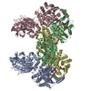 6ofcC 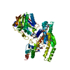 3ilvS C: citing same article ( S: Starting model for refinement |
|---|---|
| Similar structure data |


-Links
 PDBj
PDBj


- Assembly
| Deposited unit | 
| ||||||||||
|---|---|---|---|---|---|---|---|---|---|---|---|
| 1 | 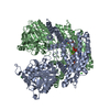
| ||||||||||
| Unit cell |
|
-Components
-Protein , 1 types, 2 molecules AB
| #1: Protein | Mass: 79481.719 Da / Num. of mol.: 2 Source method: isolated from a genetically manipulated source Source: (gene. exp.) Homo sapiens (human) / Gene: NADSYN1 / Plasmid: PI-SUMOSTAR / Cell line (production host): Sf9 / Production host:   Spodoptera frugiperda (fall armyworm) Spodoptera frugiperda (fall armyworm)References: UniProt: Q6IA69, NAD+ synthase (glutamine-hydrolysing) |
|---|
-Non-polymers , 6 types, 64 molecules 


| #2: Chemical |  Mass: 665.418 Da / Num. of mol.: 2 / Source method: obtained synthetically / Formula: C21H27N6O15P2 / Feature type: SUBJECT OF INVESTIGATION Mass: 665.418 Da / Num. of mol.: 2 / Source method: obtained synthetically / Formula: C21H27N6O15P2 / Feature type: SUBJECT OF INVESTIGATION#3: Chemical | Adenosine monophosphate Mass: 347.221 Da / Num. of mol.: 2 / Source method: obtained synthetically / Formula: C10H14N5O7P / Feature type: SUBJECT OF INVESTIGATION / Comment: AMP*YM Mass: 347.221 Da / Num. of mol.: 2 / Source method: obtained synthetically / Formula: C10H14N5O7P / Feature type: SUBJECT OF INVESTIGATION / Comment: AMP*YM#4: Chemical | Pyrophosphate Mass: 175.959 Da / Num. of mol.: 2 / Source method: obtained synthetically / Formula: H2O7P2 / Feature type: SUBJECT OF INVESTIGATION Mass: 175.959 Da / Num. of mol.: 2 / Source method: obtained synthetically / Formula: H2O7P2 / Feature type: SUBJECT OF INVESTIGATION#5: Chemical |  Mass: 24.305 Da / Num. of mol.: 2 / Source method: obtained synthetically / Formula: Mg / Feature type: SUBJECT OF INVESTIGATION Mass: 24.305 Da / Num. of mol.: 2 / Source method: obtained synthetically / Formula: Mg / Feature type: SUBJECT OF INVESTIGATION#6: Chemical | ChemComp-CL / Chloride Mass: 35.453 Da / Num. of mol.: 9 / Source method: obtained synthetically / Formula: Cl Mass: 35.453 Da / Num. of mol.: 9 / Source method: obtained synthetically / Formula: Cl#7: Water | ChemComp-HOH / | WaterMass: 18.015 Da / Num. of mol.: 47 / Source method: isolated from a natural source / Formula: H2O |
|---|
-Details
| Has ligand of interest | Y |
|---|
-Experimental details
-Experiment
| Experiment | Method: X-RAY DIFFRACTION / Number of used crystals: 1 |
|---|
- Sample preparation
| Crystal | Density Matthews: 3.51 Å3/Da / Density % sol: 64.99 % |
|---|---|
| Crystal grow | Temperature: 288 K / Method: vapor diffusion, hanging drop / pH: 7.5 Details: 0.8 M AMMONIUM SULFATE, 30% MPD, 15% GLYCEROL, 85 MM HEPES PH range: 7.5 |
-Data collection
| Diffraction | Mean temperature: 100 K / Serial crystal experiment: N |
|---|---|
| Diffraction source | Source: SYNCHROTRON / Site: SSRL  / Beamline: BL7-1 / Wavelength: 0.9795 Å / Beamline: BL7-1 / Wavelength: 0.9795 Å |
| Detector | Type: ADSC QUANTUM 315r / Detector: CCD / Date: Jun 30, 2010 / Details: MIRROR |
| Radiation | Protocol: SINGLE WAVELENGTH / Monochromatic (M) / Laue (L): M / Scattering type: x-ray |
| Radiation wavelength | Wavelength: 0.9795 Å / Relative weight: 1 |
| Reflection | Resolution: 2.84→40 Å / Num. obs: 52456 / % possible obs: 99.5 % / Redundancy: 4.1 % / Biso Wilson estimate: 71.78 Å2 / Rmerge(I) obs: 0.105 / Rsym value: 0.063 / Net I/σ(I): 13 |
| Reflection shell | Resolution: 2.84→2.91 Å / Redundancy: 4.2 % / Mean I/σ(I) obs: 1.3 / Num. unique obs: 3857 / Rsym value: 0.586 / % possible all: 99.7 |
- Processing
| Software |
| ||||||||||||||||||||||||||||||||||||||||||||||||||||||||||||||||||||||||||||||||||||||||||||||||||||||||||||||||||||||||||||||||||||||||||||
|---|---|---|---|---|---|---|---|---|---|---|---|---|---|---|---|---|---|---|---|---|---|---|---|---|---|---|---|---|---|---|---|---|---|---|---|---|---|---|---|---|---|---|---|---|---|---|---|---|---|---|---|---|---|---|---|---|---|---|---|---|---|---|---|---|---|---|---|---|---|---|---|---|---|---|---|---|---|---|---|---|---|---|---|---|---|---|---|---|---|---|---|---|---|---|---|---|---|---|---|---|---|---|---|---|---|---|---|---|---|---|---|---|---|---|---|---|---|---|---|---|---|---|---|---|---|---|---|---|---|---|---|---|---|---|---|---|---|---|---|---|---|
| Refinement | Method to determine structure: MOLECULAR REPLACEMENT Starting model: 3ILV Resolution: 2.84→37.4 Å / SU ML: 0.4285 / Cross valid method: FREE R-VALUE / σ(F): 1.33 / Phase error: 22.8257 Stereochemistry target values: GeoStd + Monomer Library + CDL v1.2
| ||||||||||||||||||||||||||||||||||||||||||||||||||||||||||||||||||||||||||||||||||||||||||||||||||||||||||||||||||||||||||||||||||||||||||||
| Solvent computation | Shrinkage radii: 0.9 Å / VDW probe radii: 1.11 Å / Solvent model: FLAT BULK SOLVENT MODEL | ||||||||||||||||||||||||||||||||||||||||||||||||||||||||||||||||||||||||||||||||||||||||||||||||||||||||||||||||||||||||||||||||||||||||||||
| Displacement parameters | Biso mean: 73.82 Å2 | ||||||||||||||||||||||||||||||||||||||||||||||||||||||||||||||||||||||||||||||||||||||||||||||||||||||||||||||||||||||||||||||||||||||||||||
| Refinement step | Cycle: LAST / Resolution: 2.84→37.4 Å
| ||||||||||||||||||||||||||||||||||||||||||||||||||||||||||||||||||||||||||||||||||||||||||||||||||||||||||||||||||||||||||||||||||||||||||||
| Refine LS restraints |
| ||||||||||||||||||||||||||||||||||||||||||||||||||||||||||||||||||||||||||||||||||||||||||||||||||||||||||||||||||||||||||||||||||||||||||||
| LS refinement shell |
|
