 Movie
Movie Controller


+ Open data

- Basic information
| Entry | Database: PDB / ID: 6lvm | ||||||
|---|---|---|---|---|---|---|---|
| Title | Crystal structure of FGFR3 in complex with pyrimidine derivative | ||||||
 Components Components | Fibroblast growth factor receptor 3 | ||||||
 Keywords Keywords | TRANSFERASE / protein kinase / SIGNALING PROTEIN | ||||||
| Function / homology |  Function and homology information Function and homology informationfibroblast growth factor receptor apoptotic signaling pathway / t(4;14) translocations of FGFR3 / negative regulation of developmental growth / Signaling by FGFR3 fusions in cancer / bone maturation / chondrocyte proliferation / endochondral bone growth / FGFR3b ligand binding and activation / Signaling by activated point mutants of FGFR3 / FGFR3c ligand binding and activation ...fibroblast growth factor receptor apoptotic signaling pathway / t(4;14) translocations of FGFR3 / negative regulation of developmental growth / Signaling by FGFR3 fusions in cancer / bone maturation / chondrocyte proliferation / endochondral bone growth / FGFR3b ligand binding and activation / Signaling by activated point mutants of FGFR3 / FGFR3c ligand binding and activation / Phospholipase C-mediated cascade; FGFR3 / fibroblast growth factor receptor activity / endochondral ossification / positive regulation of phospholipase activity / bone morphogenesis / PI-3K cascade:FGFR3 / fibroblast growth factor binding / bone mineralization / cell surface receptor signaling pathway via JAK-STAT / PI3K Cascade / fibroblast growth factor receptor signaling pathway / chondrocyte differentiation / SHC-mediated cascade:FGFR3 / FRS-mediated FGFR3 signaling / positive regulation of tyrosine phosphorylation of STAT protein / transport vesicle / Signaling by FGFR3 in disease / skeletal system development / Negative regulation of FGFR3 signaling / cell surface receptor protein tyrosine kinase signaling pathway / receptor protein-tyrosine kinase / Constitutive Signaling by Aberrant PI3K in Cancer / MAPK cascade / PIP3 activates AKT signaling / cell-cell signaling / PI5P, PP2A and IER3 Regulate PI3K/AKT Signaling / RAF/MAP kinase cascade / protein tyrosine kinase activity / positive regulation of MAPK cascade / positive regulation of phosphatidylinositol 3-kinase/protein kinase B signal transduction / positive regulation of ERK1 and ERK2 cascade / receptor complex / phosphorylation / positive regulation of cell population proliferation / Golgi apparatus / cell surface / endoplasmic reticulum / extracellular region / ATP binding / identical protein binding / plasma membraneSimilarity search - Function | ||||||
| Biological species |  Homo sapiens (human) Homo sapiens (human) | ||||||
| Method | X-RAY DIFFRACTION / SYNCHROTRON / MOLECULAR REPLACEMENT / Resolution: 2.53 Å | ||||||
 Authors Authors | Echizen, Y. / Tateishi, Y. / Amano, Y. | ||||||
 Citation Citation | Journal: Bioorg.Med.Chem. / Year: 2020 Title: Structure-based drug design of 1,3,5-triazine and pyrimidine derivatives as novel FGFR3 inhibitors with high selectivity over VEGFR2. Authors: Kuriwaki, I. / Kameda, M. / Hisamichi, H. / Kikuchi, S. / Iikubo, K. / Kawamoto, Y. / Moritomo, H. / Kondoh, Y. / Amano, Y. / Tateishi, Y. / Echizen, Y. / Iwai, Y. / Noda, A. / Tomiyama, H. ...Authors: Kuriwaki, I. / Kameda, M. / Hisamichi, H. / Kikuchi, S. / Iikubo, K. / Kawamoto, Y. / Moritomo, H. / Kondoh, Y. / Amano, Y. / Tateishi, Y. / Echizen, Y. / Iwai, Y. / Noda, A. / Tomiyama, H. / Suzuki, T. / Hirano, M. | ||||||
| History |
|
- Structure visualization
| Structure viewer | Molecule: MolmilJmol/JSmol |
|---|
- Downloads & links
-Download
| PDBx/mmCIF format | 6lvm.cif.gz | 78.3 KB | Display | PDBx/mmCIF format |
|---|---|---|---|---|
| PDB format | pdb6lvm.ent.gz | 54.7 KB | Display | PDB format |
| PDBx/mmJSON format | 6lvm.json.gz | Tree view | PDBx/mmJSON format | |
| Others |  Other downloads Other downloads |
-Validation report
| Arichive directory | https://data.pdbj.org/pub/pdb/validation_reports/lv/6lvmftp://data.pdbj.org/pub/pdb/validation_reports/lv/6lvm | HTTPS FTP |
|---|
-Related structure data
| Related structure data |  6lvkC  6lvlC  3b2tS C: citing same article ( S: Starting model for refinement |
|---|---|
| Similar structure data |


-Links
 PDBj
PDBj


- Assembly
| Deposited unit | 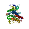
| ||||||||
|---|---|---|---|---|---|---|---|---|---|
| 1 |
| ||||||||
| Unit cell |
|
-Components
| #1: Protein | / FGFR-3 Mass: 35664.734 Da / Num. of mol.: 1 Source method: isolated from a genetically manipulated source Source: (gene. exp.) Homo sapiens (human) / Gene: FGFR3, JTK4 / Production host:  Escherichia coli (E. coli) Escherichia coli (E. coli)References: UniProt: P22607, receptor protein-tyrosine kinase |
|---|---|
| #2: Chemical | ChemComp-EVR /   Mass: 744.946 Da / Num. of mol.: 1 / Source method: obtained synthetically / Formula: C39H52N8O5S / Feature type: SUBJECT OF INVESTIGATION Mass: 744.946 Da / Num. of mol.: 1 / Source method: obtained synthetically / Formula: C39H52N8O5S / Feature type: SUBJECT OF INVESTIGATION |
| #3: Water | ChemComp-HOH / Water Mass: 18.015 Da / Num. of mol.: 31 / Source method: isolated from a natural source / Formula: H2O Mass: 18.015 Da / Num. of mol.: 31 / Source method: isolated from a natural source / Formula: H2O |
| Has ligand of interest | Y |
-Experimental details
-Experiment
| Experiment | Method: X-RAY DIFFRACTION / Number of used crystals: 1 |
|---|
- Sample preparation
| Crystal | Density Matthews: 2.27 Å3/Da / Density % sol: 45.79 % |
|---|---|
| Crystal grow | Temperature: 296 K / Method: vapor diffusion, sitting drop / pH: 5 / Details: Citric Acid, PEG 8000 |
-Data collection
| Diffraction | Mean temperature: 90 K / Serial crystal experiment: N | |||||||||||||||||||||||||||||||||||||||||||||||||||||||||||||||||||||||||||||||||||||||||||||||||||||||||||||||||||||||||||||||||||||||||||||||||||
|---|---|---|---|---|---|---|---|---|---|---|---|---|---|---|---|---|---|---|---|---|---|---|---|---|---|---|---|---|---|---|---|---|---|---|---|---|---|---|---|---|---|---|---|---|---|---|---|---|---|---|---|---|---|---|---|---|---|---|---|---|---|---|---|---|---|---|---|---|---|---|---|---|---|---|---|---|---|---|---|---|---|---|---|---|---|---|---|---|---|---|---|---|---|---|---|---|---|---|---|---|---|---|---|---|---|---|---|---|---|---|---|---|---|---|---|---|---|---|---|---|---|---|---|---|---|---|---|---|---|---|---|---|---|---|---|---|---|---|---|---|---|---|---|---|---|---|---|---|
| Diffraction source | Source: SYNCHROTRON / Site: Photon Factory  / Beamline: AR-NE3A / Wavelength: 1 Å / Beamline: AR-NE3A / Wavelength: 1 Å | |||||||||||||||||||||||||||||||||||||||||||||||||||||||||||||||||||||||||||||||||||||||||||||||||||||||||||||||||||||||||||||||||||||||||||||||||||
| Detector | Type: ADSC QUANTUM 270 / Detector: CCD / Date: Dec 13, 2010 | |||||||||||||||||||||||||||||||||||||||||||||||||||||||||||||||||||||||||||||||||||||||||||||||||||||||||||||||||||||||||||||||||||||||||||||||||||
| Radiation | Protocol: SINGLE WAVELENGTH / Monochromatic (M) / Laue (L): M / Scattering type: x-ray | |||||||||||||||||||||||||||||||||||||||||||||||||||||||||||||||||||||||||||||||||||||||||||||||||||||||||||||||||||||||||||||||||||||||||||||||||||
| Radiation wavelength | Wavelength: 1 Å / Relative weight: 1 | |||||||||||||||||||||||||||||||||||||||||||||||||||||||||||||||||||||||||||||||||||||||||||||||||||||||||||||||||||||||||||||||||||||||||||||||||||
| Reflection | Resolution: 2.53→50 Å / Num. obs: 10740 / % possible obs: 99.4 % / Redundancy: 3.6 % / Rmerge(I) obs: 0.073 / Χ2: 2.206 / Net I/σ(I): 13.9 | |||||||||||||||||||||||||||||||||||||||||||||||||||||||||||||||||||||||||||||||||||||||||||||||||||||||||||||||||||||||||||||||||||||||||||||||||||
| Reflection shell |
|
- Processing
| Software |
| |||||||||||||||||||||||||||||||||||||||||||||
|---|---|---|---|---|---|---|---|---|---|---|---|---|---|---|---|---|---|---|---|---|---|---|---|---|---|---|---|---|---|---|---|---|---|---|---|---|---|---|---|---|---|---|---|---|---|---|
| Refinement | Method to determine structure: MOLECULAR REPLACEMENT Starting model: 3B2T Resolution: 2.53→29.36 Å / Cor.coef. Fo:Fc: 0.948 / Cor.coef. Fo:Fc free: 0.896 / SU B: 12.568 / SU ML: 0.27 / Cross valid method: THROUGHOUT / σ(F): 0 / ESU R: 0.996 / ESU R Free: 0.347 / Details: U VALUES : REFINED INDIVIDUALLY
| |||||||||||||||||||||||||||||||||||||||||||||
| Solvent computation | Ion probe radii: 0.8 Å / Shrinkage radii: 0.8 Å / VDW probe radii: 1.2 Å | |||||||||||||||||||||||||||||||||||||||||||||
| Displacement parameters | Biso max: 131.43 Å2 / Biso mean: 56.476 Å2 / Biso min: 26.48 Å2
| |||||||||||||||||||||||||||||||||||||||||||||
| Refinement step | Cycle: final / Resolution: 2.53→29.36 Å
| |||||||||||||||||||||||||||||||||||||||||||||
| Refine LS restraints |
| |||||||||||||||||||||||||||||||||||||||||||||
| LS refinement shell | Resolution: 2.53→2.59 Å / Rfactor Rfree error: 0
|
