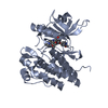
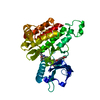
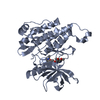
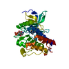
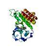
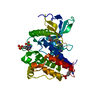
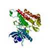
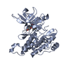
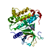
[English] 日本語
Yorodumi
- PDB-6xvj: Crystal structure of the KDR (VEGFR2) kinase domain in complex wi... -

+
Open data


ID or keywords:

Loading...

-
Basic information

Entry
Database: PDB / ID: 6xvj
TitleCrystal structure of the KDR (VEGFR2) kinase domain in complex with a type-II inhibitor
ComponentsVascular endothelial growth factor receptor 2VEGF receptor
KeywordsSIGNALING PROTEIN / type II kinase inhibitor / structure-based drug design
Function / homology
Function and homology information


blood vessel endothelial cell differentiation / cellular response to hydrogen sulfide / regulation of bone development / Signaling by membrane-tethered fusions of PDGFRA or PDGFRB / vascular endothelial growth factor binding / Neurophilin interactions with VEGF and VEGFR / endothelium development / vascular endothelial growth factor receptor-2 signaling pathway / VEGF binds to VEGFR leading to receptor dimerization / endocardium development ...blood vessel endothelial cell differentiation / cellular response to hydrogen sulfide / regulation of bone development / Signaling by membrane-tethered fusions of PDGFRA or PDGFRB / vascular endothelial growth factor binding / Neurophilin interactions with VEGF and VEGFR / endothelium development / vascular endothelial growth factor receptor-2 signaling pathway / VEGF binds to VEGFR leading to receptor dimerization / endocardium development / vascular wound healing / vascular endothelial growth factor receptor activity / regulation of hematopoietic progenitor cell differentiation / post-embryonic camera-type eye morphogenesis / endothelial cell differentiation / mesenchymal cell proliferation / positive regulation of vasculogenesis / lymph vessel development / positive regulation of BMP signaling pathway / surfactant homeostasis / cell migration involved in sprouting angiogenesis / anchoring junction / epithelial cell maturation / positive regulation of positive chemotaxis / embryonic hemopoiesis / vascular endothelial growth factor signaling pathway / positive regulation of endothelial cell chemotaxis / positive regulation of mesenchymal cell proliferation / positive regulation of mitochondrial depolarization / branching involved in blood vessel morphogenesis / positive regulation of cell migration involved in sprouting angiogenesis / positive regulation of mitochondrial fission / lung alveolus development / positive regulation of stem cell proliferation / positive regulation of nitric-oxide synthase biosynthetic process / sorting endosome / growth factor binding / positive regulation of focal adhesion assembly / regulation of MAPK cascade / semaphorin-plexin signaling pathway / positive regulation of macroautophagy / : / positive regulation of blood vessel endothelial cell migration / cellular response to vascular endothelial growth factor stimulus / cell fate commitment / calcium ion homeostasis / Integrin cell surface interactions / vasculogenesis / vascular endothelial growth factor receptor signaling pathway / coreceptor activity / negative regulation of endothelial cell apoptotic process / peptidyl-tyrosine autophosphorylation / ovarian follicle development / positive regulation of endothelial cell proliferation / transmembrane receptor protein tyrosine kinase activity / positive regulation of endothelial cell migration / VEGFR2 mediated cell proliferation / epithelial cell proliferation / stem cell proliferation / cell surface receptor protein tyrosine kinase signaling pathway / Hsp90 protein binding / receptor protein-tyrosine kinase / VEGFA-VEGFR2 Pathway / peptidyl-tyrosine phosphorylation / positive regulation of angiogenesis / cell migration / integrin binding / cell junction / regulation of cell shape / protein tyrosine kinase activity / angiogenesis / negative regulation of neuron apoptotic process / positive regulation of MAPK cascade / protein autophosphorylation / positive regulation of phosphatidylinositol 3-kinase/protein kinase B signal transduction / early endosome / positive regulation of ERK1 and ERK2 cascade / receptor complex / endosome / positive regulation of cell migration / cadherin binding / membrane raft / positive regulation of protein phosphorylation / external side of plasma membrane / negative regulation of gene expression / positive regulation of cell population proliferation / negative regulation of apoptotic process / Golgi apparatus / endoplasmic reticulum / extracellular region / ATP binding / identical protein binding / nucleus / plasma membrane
Similarity search - Function
Vascular endothelial growth factor receptor 2 (VEGFR2) / VEGFR-2, transmembrane domain / VEGFR-2 Transmembrane domain / Vascular endothelial growth factor receptor 1-like, Ig-like domain / Tyrosine-protein kinase, receptor class III, conserved site / Receptor tyrosine kinase class III signature. / Immunoglobulin domain / Immunoglobulin / Immunoglobulin domain / Immunoglobulin I-set ...Vascular endothelial growth factor receptor 2 (VEGFR2) / VEGFR-2, transmembrane domain / VEGFR-2 Transmembrane domain / Vascular endothelial growth factor receptor 1-like, Ig-like domain / Tyrosine-protein kinase, receptor class III, conserved site / Receptor tyrosine kinase class III signature. / Immunoglobulin domain / Immunoglobulin / Immunoglobulin domain / Immunoglobulin I-set / Immunoglobulin I-set domain / Immunoglobulin subtype 2 / Immunoglobulin C-2 Type / Tyrosine-protein kinase, catalytic domain / Tyrosine kinase, catalytic domain / Tyrosine protein kinases specific active-site signature. / Immunoglobulin subtype / Immunoglobulin / Tyrosine-protein kinase, active site / Protein tyrosine and serine/threonine kinase / Serine-threonine/tyrosine-protein kinase, catalytic domain / Ig-like domain profile. / Immunoglobulin-like domain / Immunoglobulin-like domain superfamily / Protein kinase, ATP binding site / Protein kinases ATP-binding region signature. / Immunoglobulin-like fold / Protein kinase domain profile. / Protein kinase domain / Protein kinase-like domain superfamily
Similarity search - Domain/homology
Chem-O35 / Vascular endothelial growth factor receptor 2
Similarity search - Component
Biological speciesHomo sapiens (human)
MethodX-RAY DIFFRACTION / SYNCHROTRON / MOLECULAR REPLACEMENT / molecular replacement / Resolution: 1.78 Å
AuthorsSchimpl, M. / McAuley, K. / Hoyt, E.A. / Thomas, M. / Bodnarchuk, M.S. / Lewis, H.J. / Barratt, D. / Deery, M.J. / Ogg, D.J. / Bernardes, G.J.L. ...Schimpl, M. / McAuley, K. / Hoyt, E.A. / Thomas, M. / Bodnarchuk, M.S. / Lewis, H.J. / Barratt, D. / Deery, M.J. / Ogg, D.J. / Bernardes, G.J.L. / Ward, R.A. / Kettle, J.G. / Waring, M.J.
CitationJournal: J.Am.Chem.Soc. / Year: 2020
Title: Alkynyl Benzoxazines and Dihydroquinazolines as Cysteine Targeting Covalent Warheads and Their Application in Identification of Selective Irreversible Kinase Inhibitors.
Authors: McAulay, K. / Hoyt, E.A. / Thomas, M. / Schimpl, M. / Bodnarchuk, M.S. / Lewis, H.J. / Barratt, D. / Bhavsar, D. / Robinson, D.M. / Deery, M.J. / Ogg, D.J. / Bernardes, G.J.L. / Ward, R.A. / ...Authors: McAulay, K. / Hoyt, E.A. / Thomas, M. / Schimpl, M. / Bodnarchuk, M.S. / Lewis, H.J. / Barratt, D. / Bhavsar, D. / Robinson, D.M. / Deery, M.J. / Ogg, D.J. / Bernardes, G.J.L. / Ward, R.A. / Waring, M.J. / Kettle, J.G.
History
DepositionJan 22, 2020Deposition site: PDBE / Processing site: PDBE
Revision 1.0May 27, 2020Provider: repository / Type: Initial release
Revision 1.1Jun 17, 2020Group: Database references / Category: citation / citation_author
Item: _citation.journal_volume / _citation.page_first ..._citation.journal_volume / _citation.page_first / _citation.page_last / _citation_author.name
Revision 1.2May 1, 2024Group: Data collection / Database references / Refinement description
Category: chem_comp_atom / chem_comp_bond ...chem_comp_atom / chem_comp_bond / database_2 / pdbx_initial_refinement_model
Item: _database_2.pdbx_DOI / _database_2.pdbx_database_accession

-
Structure visualization

Structure viewerMolecule:
MolmilJmol/JSmol

Downloads & links

-
Assembly

Deposited unit
A: Vascular endothelial growth factor receptor 2
hetero molecules


Theoretical massNumber of molelcules
Total (without water)37,4762
Polymers36,9901
Non-polymers4871
Water3,693205
1


  • Idetical with deposited unit
  • defined by author&software
  • Evidence: gel filtration
TypeNameSymmetry operationNumber
identity operation1_555x,y,z1
Buried area0 Å2
ΔGint0 kcal/mol
Surface area15180 Å2
MethodPISA
Unit cell
Length a, b, c (Å)51.724, 56.257, 60.260
Angle α, β, γ (deg.)90.000, 110.400, 90.000
Int Tables number4
Space group name H-MP1211

-
Components

#1: Protein Vascular endothelial growth factor receptor 2 / VEGF receptor / VEGFR-2 / Fetal liver kinase 1 / FLK-1 / Kinase insert domain receptor / KDR / Protein-tyrosine ...VEGFR-2 / Fetal liver kinase 1 / FLK-1 / Kinase insert domain receptor / KDR / Protein-tyrosine kinase receptor flk-1


Mass: 36989.609 Da / Num. of mol.: 1 / Fragment: protein kinase domain (807-1171 del[940-989]) / Mutation: E990V
Source method: isolated from a genetically manipulated source
Source: (gene. exp.) Homo sapiens (human) / Gene: KDR, FLK1, VEGFR2 / Plasmid: pFastBac1 / Cell line (production host): Sf21 / Production host: Spodoptera frugiperda (fall armyworm)
References: UniProt: P35968, receptor protein-tyrosine kinase
#2: Chemical ChemComp-O35 / ~{N}-[3-[(dimethylamino)methyl]-5-methyl-phenyl]-2-[3-methoxy-5-(7-methoxyquinolin-4-yl)oxy-pyridin-2-yl]ethanamide


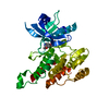
Mass: 486.562 Da / Num. of mol.: 1 / Source method: obtained synthetically / Formula: C28H30N4O4 / Feature type: SUBJECT OF INVESTIGATION
#3: Water ChemComp-HOH / water / Water


Mass: 18.015 Da / Num. of mol.: 205 / Source method: isolated from a natural source / Formula: H2O
Has ligand of interestY

-
Experimental details

-
Experiment

ExperimentMethod: X-RAY DIFFRACTION / Number of used crystals: 1

-
Sample preparation

CrystalDensity Matthews: 2.22 Å3/Da / Density % sol: 44.63 % / Mosaicity: 0.42 °
Crystal growTemperature: 293 K / Method: vapor diffusion / pH: 8.5
Details: 25 % PEG550MME, 0.1 M NaCl, 0.1 M TRIS buffer pH 8.5

-
Data collection

DiffractionMean temperature: 100 K / Serial crystal experiment: N
Diffraction sourceSource: SYNCHROTRON / Site: Diamond / Beamline: I03 / Wavelength: 0.97626 Å
DetectorType: DECTRIS PILATUS 6M / Detector: PIXEL / Date: Aug 1, 2015
RadiationProtocol: SINGLE WAVELENGTH / Monochromatic (M) / Laue (L): M / Scattering type: x-ray
Radiation wavelengthWavelength: 0.97626 Å / Relative weight: 1
ReflectionResolution: 1.78→56.48 Å / Num. obs: 29228 / % possible obs: 93.5 % / Redundancy: 3.2 % / Biso Wilson estimate: 30.67 Å2 / CC1/2: 0.999 / Rmerge(I) obs: 0.042 / Rpim(I) all: 0.027 / Rrim(I) all: 0.05 / Net I/σ(I): 15.4 / Num. measured all: 93735
Reflection shell

Diffraction-ID: 1

Resolution (Å)Redundancy (%)Rmerge(I) obsNum. measured allNum. unique obsCC1/2Rpim(I) allRrim(I) allNet I/σ(I) obs% possible all
1.78-1.883.30.491477845330.8010.3190.5872.399.9
5.63-56.483.10.025323510330.9980.0170.033999.2

-
Phasing

PhasingMethod: molecular replacement

-
Processing

Software
NameVersionClassificationNB
XDSdata reduction
Aimless0.5.12data scaling
AMoREphasing
BUSTERrefinement
PDB_EXTRACT3.25data extraction
RefinementMethod to determine structure: MOLECULAR REPLACEMENT
Starting model: internal model

Resolution: 1.78→48.48 Å / Cor.coef. Fo:Fc: 0.955 / Cor.coef. Fo:Fc free: 0.95 / SU R Cruickshank DPI: 0.123 / Cross valid method: THROUGHOUT / σ(F): 0 / SU R Blow DPI: 0.126 / SU Rfree Blow DPI: 0.119 / SU Rfree Cruickshank DPI: 0.118
RfactorNum. reflection% reflectionSelection details
Rfree0.215 1415 4.84 %RANDOM
Rwork0.178 ---
obs0.18 29206 93.5 %-
Displacement parametersBiso max: 119.43 Å2 / Biso mean: 36.22 Å2 / Biso min: 16.46 Å2
Baniso -1Baniso -2Baniso -3
1-3.8222 Å20 Å2-0.1467 Å2
2---0.086 Å20 Å2
3----3.7362 Å2
Refine analyzeLuzzati coordinate error obs: 0.22 Å
Refinement stepCycle: final / Resolution: 1.78→48.48 Å
ProteinNucleic acidLigandSolventTotal
Num. atoms2443 0 36 205 2684
Biso mean--24.57 43.17 -
Num. residues----305
Refine LS restraints
Refine-IDTypeNumberRestraint functionWeightDev ideal
X-RAY DIFFRACTIONt_dihedral_angle_d888SINUSOIDAL2
X-RAY DIFFRACTIONt_trig_c_planes
X-RAY DIFFRACTIONt_gen_planes422HARMONIC5
X-RAY DIFFRACTIONt_it2543HARMONIC20
X-RAY DIFFRACTIONt_nbd
X-RAY DIFFRACTIONt_improper_torsion
X-RAY DIFFRACTIONt_pseud_angle
X-RAY DIFFRACTIONt_chiral_improper_torsion314SEMIHARMONIC5
X-RAY DIFFRACTIONt_sum_occupancies
X-RAY DIFFRACTIONt_utility_distance
X-RAY DIFFRACTIONt_utility_angle
X-RAY DIFFRACTIONt_utility_torsion
X-RAY DIFFRACTIONt_ideal_dist_contact3133SEMIHARMONIC4
X-RAY DIFFRACTIONt_bond_d2543HARMONIC20.01
X-RAY DIFFRACTIONt_angle_deg3440HARMONIC20.99
X-RAY DIFFRACTIONt_omega_torsion3.48
X-RAY DIFFRACTIONt_other_torsion17.73
LS refinement shellResolution: 1.78→1.79 Å / Rfactor Rfree error: 0 / Total num. of bins used: 50
RfactorNum. reflection% reflection
Rfree0.3372 27 4.62 %
Rwork0.2086 558 -
all0.2143 585 -
obs--99.47 %
Refinement TLS params.Method: refined / Origin x: 21.3629 Å / Origin y: -0.5065 Å / Origin z: 12.1668 Å
111213212223313233
T-0.0805 Å20.0003 Å20.0163 Å2--0.0564 Å2-0.0068 Å2---0.0858 Å2
L1.2963 °2-0.1197 °20.1831 °2-1.044 °2-0.334 °2--1.182 °2
S-0.0131 Å °-0.165 Å °-0.0181 Å °0.1304 Å °-0.0203 Å °-0.0104 Å °-0.0523 Å °0.1959 Å °0.0334 Å °
Refinement TLS groupSelection details: { A|* }

+
About Yorodumi

-
News

-
Feb 9, 2022. New format data for meta-information of EMDB entries

New format data for meta-information of EMDB entries

  • Version 3 of the EMDB header file is now the official format.
  • The previous official version 1.9 will be removed from the archive.

Related info.:EMDB header

External links:wwPDB to switch to version 3 of the EMDB data model

-
Aug 12, 2020. Covid-19 info

Covid-19 info

URL: https://pdbj.org/emnavi/covid19.php

New page: Covid-19 featured information page in EM Navigator.

Related info.:Covid-19 info / Mar 5, 2020. Novel coronavirus structure data

+
Mar 5, 2020. Novel coronavirus structure data

Novel coronavirus structure data

Related info.:Yorodumi Speices / Aug 12, 2020. Covid-19 info

External links:COVID-19 featured content - PDBj / Molecule of the Month (242):Coronavirus Proteases

+
Jan 31, 2019. EMDB accession codes are about to change! (news from PDBe EMDB page)

EMDB accession codes are about to change! (news from PDBe EMDB page)

  • The allocation of 4 digits for EMDB accession codes will soon come to an end. Whilst these codes will remain in use, new EMDB accession codes will include an additional digit and will expand incrementally as the available range of codes is exhausted. The current 4-digit format prefixed with “EMD-” (i.e. EMD-XXXX) will advance to a 5-digit format (i.e. EMD-XXXXX), and so on. It is currently estimated that the 4-digit codes will be depleted around Spring 2019, at which point the 5-digit format will come into force.
  • The EM Navigator/Yorodumi systems omit the EMD- prefix.

Related info.:Q: What is EMD? / ID/Accession-code notation in Yorodumi/EM Navigator

External links:EMDB Accession Codes are Changing Soon! / Contact to PDBj

+
Jul 12, 2017. Major update of PDB

Major update of PDB

  • wwPDB released updated PDB data conforming to the new PDBx/mmCIF dictionary.
  • This is a major update changing the version number from 4 to 5, and with Remediation, in which all the entries are updated.
  • In this update, many items about electron microscopy experimental information are reorganized (e.g. em_software).
  • Now, EM Navigator and Yorodumi are based on the updated data.

External links:wwPDB Remediation / Enriched Model Files Conforming to OneDep Data Standards Now Available in the PDB FTP Archive

-
Yorodumi

Thousand views of thousand structures

  • Yorodumi is a browser for structure data from EMDB, PDB, SASBDB, etc.
  • This page is also the successor to EM Navigator detail page, and also detail information page/front-end page for Omokage search.
  • The word "yorodu" (or yorozu) is an old Japanese word meaning "ten thousand". "mi" (miru) is to see.

Related info.:EMDB / PDB / SASBDB / Comparison of 3 databanks / Yorodumi Search / Aug 31, 2016. New EM Navigator & Yorodumi / Yorodumi Papers / Jmol/JSmol / Function and homology information / Changes in new EM Navigator and Yorodumi

Read more