 Movie
Movie Controller

[English] 日本語
 Yorodumi
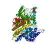
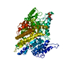
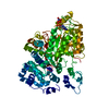
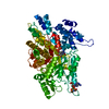
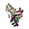
- PDB-6l3r: Crystal structure of Ribonucleotide reductase R1 subunit, RRM1 in... -
+ Open data

- Basic information
| Entry | Database: PDB / ID: 6l3r | ||||||
|---|---|---|---|---|---|---|---|
| Title | Crystal structure of Ribonucleotide reductase R1 subunit, RRM1 in complex with 4-bromo-N-((1S,2R)-2-(naphthalen-1-yl)-1-(5-oxo-4,5-dihydro-1,3,4-oxadiazol-2-yl)propyl)benzenesulfonamide | ||||||
 Components Components | Ribonucleoside-diphosphate reductase large subunit Ribonucleotide reductase Ribonucleotide reductase | ||||||
 Keywords Keywords | OXIDOREDUCTASE | ||||||
| Function / homology |  Function and homology informationribonucleoside-diphosphate reductase activity / pyrimidine nucleobase metabolic process / cell proliferation in forebrain / ribonucleoside diphosphate metabolic process / positive regulation of G0 to G1 transition / mitochondrial DNA replication / 2'-deoxyribonucleotide biosynthetic process / ribonucleoside-diphosphate reductase complex / ribonucleoside-diphosphate reductase / ribonucleoside-diphosphate reductase activity, thioredoxin disulfide as acceptor ...ribonucleoside-diphosphate reductase activity / pyrimidine nucleobase metabolic process / cell proliferation in forebrain / ribonucleoside diphosphate metabolic process / positive regulation of G0 to G1 transition / mitochondrial DNA replication / 2'-deoxyribonucleotide biosynthetic process / ribonucleoside-diphosphate reductase complex / ribonucleoside-diphosphate reductase / ribonucleoside-diphosphate reductase activity, thioredoxin disulfide as acceptor / Interconversion of nucleotide di- and triphosphates / deoxyribonucleotide biosynthetic process / protein heterotetramerization / response to ionizing radiation / DNA synthesis involved in DNA repair / positive regulation of G1/S transition of mitotic cell cycle / positive regulation of G2/M transition of mitotic cell cycle / cell projection / male gonad development / disordered domain specific binding / retina development in camera-type eye / nuclear envelope / DNA repair / neuronal cell body / mitochondrion / ATP binding / identical protein binding / cytosol Function and homology informationribonucleoside-diphosphate reductase activity / pyrimidine nucleobase metabolic process / cell proliferation in forebrain / ribonucleoside diphosphate metabolic process / positive regulation of G0 to G1 transition / mitochondrial DNA replication / 2'-deoxyribonucleotide biosynthetic process / ribonucleoside-diphosphate reductase complex / ribonucleoside-diphosphate reductase / ribonucleoside-diphosphate reductase activity, thioredoxin disulfide as acceptor ...ribonucleoside-diphosphate reductase activity / pyrimidine nucleobase metabolic process / cell proliferation in forebrain / ribonucleoside diphosphate metabolic process / positive regulation of G0 to G1 transition / mitochondrial DNA replication / 2'-deoxyribonucleotide biosynthetic process / ribonucleoside-diphosphate reductase complex / ribonucleoside-diphosphate reductase / ribonucleoside-diphosphate reductase activity, thioredoxin disulfide as acceptor / Interconversion of nucleotide di- and triphosphates / deoxyribonucleotide biosynthetic process / protein heterotetramerization / response to ionizing radiation / DNA synthesis involved in DNA repair / positive regulation of G1/S transition of mitotic cell cycle / positive regulation of G2/M transition of mitotic cell cycle / cell projection / male gonad development / disordered domain specific binding / retina development in camera-type eye / nuclear envelope / DNA repair / neuronal cell body / mitochondrion / ATP binding / identical protein binding / cytosolSimilarity search - Function | ||||||
| Biological species |  Homo sapiens (human) Homo sapiens (human) | ||||||
| Method | X-RAY DIFFRACTION / SYNCHROTRON / MOLECULAR REPLACEMENT / Resolution: 2 Å | ||||||
 Authors Authors | Miyahara, S. / Chong, K.T. / Suzuki, T. | ||||||
 Citation Citation | Journal: To be published Title: TAS1553, a novel small molecule ribonucleotide reductase (RNR) subunit interaction inhibitor, displays remarkable anti-tumor activity Authors: Miyahara, S. / Hara, S. / Chong, K.T. / Suzuki, T. / Ogino, Y. / Hoshino, T. / Tsukioka, S. / Yano, W. / Suzuki, M. / Otsu, Y. / Yonekura, T. / Ito, S. / Terasaka, M. / Suzuki, T. / Hashimoto, A. | ||||||
| History |
|
- Structure visualization
| Structure viewer | Molecule: MolmilJmol/JSmol |
|---|
- Downloads & links
-Download
| PDBx/mmCIF format | 6l3r.cif.gz | 282.5 KB | Display | PDBx/mmCIF format |
|---|---|---|---|---|
| PDB format | pdb6l3r.ent.gz | 223.5 KB | Display | PDB format |
| PDBx/mmJSON format | 6l3r.json.gz | Tree view | PDBx/mmJSON format | |
| Others |  Other downloads Other downloads |
-Validation report
| Arichive directory | https://data.pdbj.org/pub/pdb/validation_reports/l3/6l3rftp://data.pdbj.org/pub/pdb/validation_reports/l3/6l3r | HTTPS FTP |
|---|
-Related structure data
| Related structure data | 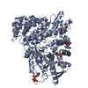 6l7lC 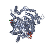 6lkmC 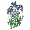 2wghS S: Starting model for refinement C: citing same article ( |
|---|---|
| Similar structure data |


-Links
 PDBj
PDBj

- Assembly
| Deposited unit | 
| ||||||||
|---|---|---|---|---|---|---|---|---|---|
| 1 | 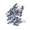
| ||||||||
| 2 | 
| ||||||||
| Unit cell |
|
-Components
-Protein , 1 types, 2 molecules AE
| #1: Protein | Ribonucleotide reductase / Ribonucleotide reductase R1 subunit Mass: 76189.859 Da / Num. of mol.: 2 Source method: isolated from a genetically manipulated source Source: (gene. exp.) Homo sapiens (human) / Gene: RRM1, RR1 / Plasmid: pET28a / Production host:  Escherichia coli BL21(DE3) (bacteria) / Strain (production host): BL21(DE3) Escherichia coli BL21(DE3) (bacteria) / Strain (production host): BL21(DE3)References: UniProt: P23921, ribonucleoside-diphosphate reductase |
|---|
-Non-polymers , 6 types, 333 molecules 


| #2: Chemical | Thymidine triphosphate Mass: 482.168 Da / Num. of mol.: 2 / Source method: obtained synthetically / Formula: C10H17N2O14P3 Mass: 482.168 Da / Num. of mol.: 2 / Source method: obtained synthetically / Formula: C10H17N2O14P3#3: Chemical |  Mass: 24.305 Da / Num. of mol.: 2 / Source method: obtained synthetically / Formula: Mg Mass: 24.305 Da / Num. of mol.: 2 / Source method: obtained synthetically / Formula: Mg#4: Chemical |  Mass: 488.354 Da / Num. of mol.: 2 / Source method: obtained synthetically / Formula: C21H18BrN3O4S / Feature type: SUBJECT OF INVESTIGATION Mass: 488.354 Da / Num. of mol.: 2 / Source method: obtained synthetically / Formula: C21H18BrN3O4S / Feature type: SUBJECT OF INVESTIGATION#5: Chemical | Acetate Mass: 59.044 Da / Num. of mol.: 3 / Source method: obtained synthetically / Formula: C2H3O2 Mass: 59.044 Da / Num. of mol.: 3 / Source method: obtained synthetically / Formula: C2H3O2#6: Chemical | ChemComp-SO4 / | Sulfate Mass: 96.063 Da / Num. of mol.: 1 / Source method: obtained synthetically / Formula: SO4 Mass: 96.063 Da / Num. of mol.: 1 / Source method: obtained synthetically / Formula: SO4#7: Water | ChemComp-HOH / | WaterMass: 18.015 Da / Num. of mol.: 323 / Source method: isolated from a natural source / Formula: H2O |
|---|
-Details
| Has ligand of interest | Y |
|---|
-Experimental details
-Experiment
| Experiment | Method: X-RAY DIFFRACTION / Number of used crystals: 1 |
|---|
- Sample preparation
| Crystal | Density Matthews: 2.5 Å3/Da / Density % sol: 50.84 % |
|---|---|
| Crystal grow | Temperature: 300 K / Method: vapor diffusion, hanging drop / Details: Sodium acetate buffer, PEG3350, Lithium sulfate |
-Data collection
| Diffraction | Mean temperature: 100 K / Serial crystal experiment: N | |||||||||||||||||||||||||||||||||||||||||||||||||||||||||||||||||||||||||||||||||||||||||||||||||||||||||||||||||||||||||
|---|---|---|---|---|---|---|---|---|---|---|---|---|---|---|---|---|---|---|---|---|---|---|---|---|---|---|---|---|---|---|---|---|---|---|---|---|---|---|---|---|---|---|---|---|---|---|---|---|---|---|---|---|---|---|---|---|---|---|---|---|---|---|---|---|---|---|---|---|---|---|---|---|---|---|---|---|---|---|---|---|---|---|---|---|---|---|---|---|---|---|---|---|---|---|---|---|---|---|---|---|---|---|---|---|---|---|---|---|---|---|---|---|---|---|---|---|---|---|---|---|---|---|
| Diffraction source | Source: SYNCHROTRON / Site: Photon Factory  / Beamline: BL-17A / Wavelength: 0.98 Å / Beamline: BL-17A / Wavelength: 0.98 Å | |||||||||||||||||||||||||||||||||||||||||||||||||||||||||||||||||||||||||||||||||||||||||||||||||||||||||||||||||||||||||
| Detector | Type: ADSC QUANTUM 270 / Detector: CCD / Date: May 10, 2013 | |||||||||||||||||||||||||||||||||||||||||||||||||||||||||||||||||||||||||||||||||||||||||||||||||||||||||||||||||||||||||
| Radiation | Protocol: SINGLE WAVELENGTH / Monochromatic (M) / Laue (L): M / Scattering type: x-ray | |||||||||||||||||||||||||||||||||||||||||||||||||||||||||||||||||||||||||||||||||||||||||||||||||||||||||||||||||||||||||
| Radiation wavelength | Wavelength: 0.98 Å / Relative weight: 1 | |||||||||||||||||||||||||||||||||||||||||||||||||||||||||||||||||||||||||||||||||||||||||||||||||||||||||||||||||||||||||
| Reflection | Resolution: 2→83.28 Å / Num. all: 101127 / Num. obs: 101127 / % possible obs: 98.9 % / Redundancy: 5.9 % / Rpim(I) all: 0.041 / Rrim(I) all: 0.102 / Rsym value: 0.093 / Net I/av σ(I): 6.2 / Net I/σ(I): 12.5 / Num. measured all: 591920 | |||||||||||||||||||||||||||||||||||||||||||||||||||||||||||||||||||||||||||||||||||||||||||||||||||||||||||||||||||||||||
| Reflection shell | Diffraction-ID: 1
|
- Processing
| Software |
| ||||||||||||||||||||||||||||||||||||||||||||||||||||||||||||
|---|---|---|---|---|---|---|---|---|---|---|---|---|---|---|---|---|---|---|---|---|---|---|---|---|---|---|---|---|---|---|---|---|---|---|---|---|---|---|---|---|---|---|---|---|---|---|---|---|---|---|---|---|---|---|---|---|---|---|---|---|---|
| Refinement | Method to determine structure: MOLECULAR REPLACEMENT Starting model: 2WGH Resolution: 2→47.92 Å / Cor.coef. Fo:Fc: 0.948 / Cor.coef. Fo:Fc free: 0.92 / SU B: 4.464 / SU ML: 0.123 / Cross valid method: THROUGHOUT / σ(F): 0 / ESU R: 0.177 / ESU R Free: 0.16 Details: HYDROGENS HAVE BEEN ADDED IN THE RIDING POSITIONS U VALUES : REFINED INDIVIDUALLY
| ||||||||||||||||||||||||||||||||||||||||||||||||||||||||||||
| Solvent computation | Ion probe radii: 0.8 Å / Shrinkage radii: 0.8 Å / VDW probe radii: 1.2 Å | ||||||||||||||||||||||||||||||||||||||||||||||||||||||||||||
| Displacement parameters | Biso max: 106.74 Å2 / Biso mean: 25.462 Å2 / Biso min: 7.6 Å2
| ||||||||||||||||||||||||||||||||||||||||||||||||||||||||||||
| Refinement step | Cycle: final / Resolution: 2→47.92 Å
| ||||||||||||||||||||||||||||||||||||||||||||||||||||||||||||
| Refine LS restraints |
| ||||||||||||||||||||||||||||||||||||||||||||||||||||||||||||
| LS refinement shell | Resolution: 2→2.052 Å / Rfactor Rfree error: 0 / Total num. of bins used: 20
|
