 Movie
Movie Controller

[English] 日本語
 Yorodumi
- PDB-6j7f: Complex of GGTaseIII, farnesyl-Ykt6 (C-terminal methylated), and GGPP -
+ Open data

- Basic information
| Entry | Database: PDB / ID: 6j7f | ||||||||||||
|---|---|---|---|---|---|---|---|---|---|---|---|---|---|
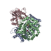
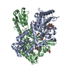
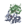
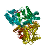
| Title | Complex of GGTaseIII, farnesyl-Ykt6 (C-terminal methylated), and GGPP | ||||||||||||
 Components Components |
| ||||||||||||
 Keywords Keywords | LIPID BINDING PROTEIN / lipid transferase | ||||||||||||
| Function / homology |  Function and homology information Function and homology informationprotein prenyltransferase activity /  protein geranylgeranyltransferase type II / protein-cysteine S-palmitoyltransferase activity / Rab-protein geranylgeranyltransferase complex / vesicle targeting / basal dendrite / Intra-Golgi traffic / Rab geranylgeranyltransferase activity / protein geranylgeranylation / SNARE complex ...protein prenyltransferase activity / protein geranylgeranyltransferase type II / protein-cysteine S-palmitoyltransferase activity / Rab-protein geranylgeranyltransferase complex / vesicle targeting / basal dendrite / Intra-Golgi traffic / Rab geranylgeranyltransferase activity / protein geranylgeranylation / SNARE complex / SNAP receptor activity / RAB geranylgeranylation / vesicle docking involved in exocytosis / apical dendrite / retrograde transport, endosome to Golgi / COPII-mediated vesicle transport / TP53 regulates transcription of several additional cell death genes whose specific roles in p53-dependent apoptosis remain uncertain / CDC42 GTPase cycle / RHOG GTPase cycle / RHOA GTPase cycle / RAC3 GTPase cycle / endoplasmic reticulum to Golgi vesicle-mediated transport / COPI-mediated anterograde transport / transport vesicle / Transferases; Acyltransferases; Transferring groups other than aminoacyl groups / RAC1 GTPase cycle / visual perception / endoplasmic reticulum-Golgi intermediate compartment membrane / protein modification process / cytoplasmic vesicle membrane / small GTPase binding / protein transport / endosome / cadherin binding / Golgi membrane / neuronal cell body / Golgi apparatus / endoplasmic reticulum / mitochondrion / zinc ion binding / plasma membrane / cytosol / cytoplasm protein geranylgeranyltransferase type II / protein-cysteine S-palmitoyltransferase activity / Rab-protein geranylgeranyltransferase complex / vesicle targeting / basal dendrite / Intra-Golgi traffic / Rab geranylgeranyltransferase activity / protein geranylgeranylation / SNARE complex ...protein prenyltransferase activity / protein geranylgeranyltransferase type II / protein-cysteine S-palmitoyltransferase activity / Rab-protein geranylgeranyltransferase complex / vesicle targeting / basal dendrite / Intra-Golgi traffic / Rab geranylgeranyltransferase activity / protein geranylgeranylation / SNARE complex / SNAP receptor activity / RAB geranylgeranylation / vesicle docking involved in exocytosis / apical dendrite / retrograde transport, endosome to Golgi / COPII-mediated vesicle transport / TP53 regulates transcription of several additional cell death genes whose specific roles in p53-dependent apoptosis remain uncertain / CDC42 GTPase cycle / RHOG GTPase cycle / RHOA GTPase cycle / RAC3 GTPase cycle / endoplasmic reticulum to Golgi vesicle-mediated transport / COPI-mediated anterograde transport / transport vesicle / Transferases; Acyltransferases; Transferring groups other than aminoacyl groups / RAC1 GTPase cycle / visual perception / endoplasmic reticulum-Golgi intermediate compartment membrane / protein modification process / cytoplasmic vesicle membrane / small GTPase binding / protein transport / endosome / cadherin binding / Golgi membrane / neuronal cell body / Golgi apparatus / endoplasmic reticulum / mitochondrion / zinc ion binding / plasma membrane / cytosol / cytoplasmSimilarity search - Function | ||||||||||||
| Biological species |  Homo sapiens (human) Homo sapiens (human) | ||||||||||||
| Method | X-RAY DIFFRACTION / SYNCHROTRON / MOLECULAR REPLACEMENT / Resolution: 2.883 Å | ||||||||||||
 Authors Authors | Goto-Ito, S. / Yamagata, A. / Sato, Y. / Fukai, S. | ||||||||||||
| Funding support |  Japan, 3items Japan, 3items
| ||||||||||||
 Citation Citation | Journal: To Be Published Title: Complex of GGTaseIII, farnesyl-Ykt6 (C-terminal methylated), and GGPP Authors: Goto-Ito, S. / Shirakawa, R. / Fukai, S. | ||||||||||||
| History |
|
- Structure visualization
| Structure viewer | Molecule: MolmilJmol/JSmol |
|---|
- Downloads & links
-Download
| PDBx/mmCIF format | 6j7f.cif.gz | 176.7 KB | Display | PDBx/mmCIF format |
|---|---|---|---|---|
| PDB format | pdb6j7f.ent.gz | 136.9 KB | Display | PDB format |
| PDBx/mmJSON format | 6j7f.json.gz | Tree view | PDBx/mmJSON format | |
| Others |  Other downloads Other downloads |
-Validation report
| Arichive directory | https://data.pdbj.org/pub/pdb/validation_reports/j7/6j7fftp://data.pdbj.org/pub/pdb/validation_reports/j7/6j7f | HTTPS FTP |
|---|
-Related structure data
| Related structure data |  6j7xC  6j6xS S: Starting model for refinement C: citing same article ( |
|---|---|
| Similar structure data |


-Links
 PDBj
PDBj


- Assembly
| Deposited unit | 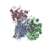
| ||||||||
|---|---|---|---|---|---|---|---|---|---|
| 1 |
| ||||||||
| Unit cell |
|
-Components
-Protein , 3 types, 3 molecules ABC
| #1: Protein | Mass: 37986.520 Da / Num. of mol.: 1 Source method: isolated from a genetically manipulated source Source: (gene. exp.) Homo sapiens (human) / Gene: PTAR1 / Production host:  Escherichia coli (E. coli) / References: UniProt: Q7Z6K3 Escherichia coli (E. coli) / References: UniProt: Q7Z6K3 |
|---|---|
| #2: Protein | Mass: 37371.723 Da / Num. of mol.: 1 Source method: isolated from a genetically manipulated source Source: (gene. exp.) Homo sapiens (human) / Gene: RABGGTB, GGTB / Production host: Escherichia coli (E. coli)References: UniProt: P53611, protein geranylgeranyltransferase type II |
| #3: Protein | Mass: 22145.174 Da / Num. of mol.: 1 Source method: isolated from a genetically manipulated source Details: farnesyl-Ykt6 (C-terminal methylated) / Source: (gene. exp.) Homo sapiens (human) / Gene: YKT6 / Production host: Escherichia coli (E. coli)References: UniProt: O15498, Transferases; Acyltransferases; Transferring groups other than aminoacyl groups |
-Non-polymers , 4 types, 4 molecules 

| #4: Chemical | ChemComp-TLA / Tartaric acid Mass: 150.087 Da / Num. of mol.: 1 / Source method: obtained synthetically / Formula: C4H6O6 Mass: 150.087 Da / Num. of mol.: 1 / Source method: obtained synthetically / Formula: C4H6O6 |
|---|---|
| #5: Chemical | ChemComp-FAR / Farnesol Mass: 206.367 Da / Num. of mol.: 1 / Source method: obtained synthetically / Formula: C15H26 / Feature type: SUBJECT OF INVESTIGATION Mass: 206.367 Da / Num. of mol.: 1 / Source method: obtained synthetically / Formula: C15H26 / Feature type: SUBJECT OF INVESTIGATION |
| #6: Chemical | ChemComp-GER /  Mass: 274.484 Da / Num. of mol.: 1 / Source method: obtained synthetically / Formula: C20H34 / Feature type: SUBJECT OF INVESTIGATION Mass: 274.484 Da / Num. of mol.: 1 / Source method: obtained synthetically / Formula: C20H34 / Feature type: SUBJECT OF INVESTIGATION |
| #7: Chemical | ChemComp-DPO / Pyrophosphate Mass: 173.943 Da / Num. of mol.: 1 / Source method: obtained synthetically / Formula: O7P2 Mass: 173.943 Da / Num. of mol.: 1 / Source method: obtained synthetically / Formula: O7P2 |
-Experimental details
-Experiment
| Experiment | Method: X-RAY DIFFRACTION / Number of used crystals: 1 |
|---|
- Sample preparation
| Crystal | Density Matthews: 3.85 Å3/Da / Density % sol: 68.06 % |
|---|---|
| Crystal grow | Temperature: 293 K / Method: vapor diffusion, sitting drop / Details: 1M Ammonium Tartrate dibasic pH 7 |
-Data collection
| Diffraction | Mean temperature: 100 K / Serial crystal experiment: N |
|---|---|
| Diffraction source | Source: SYNCHROTRON / Site: SPring-8 / Beamline: BL41XU / Wavelength: 1 Å |
| Detector | Type: DECTRIS EIGER X 16M / Detector: PIXEL / Date: Apr 12, 2018 |
| Radiation | Protocol: SINGLE WAVELENGTH / Monochromatic (M) / Laue (L): M / Scattering type: x-ray |
| Radiation wavelength | Wavelength: 1 Å / Relative weight: 1 |
| Reflection | Resolution: 2.88→50 Å / Num. obs: 35109 / % possible obs: 99.9 % / Redundancy: 16.2 % / Rsym value: 0.136 / Net I/σ(I): 12.3 |
| Reflection shell | Resolution: 2.88→2.93 Å / Redundancy: 9.8 % / Mean I/σ(I) obs: 2.1 / Num. unique obs: 1721 / Rsym value: 0.512 / % possible all: 99.9 |
- Processing
| Software |
| |||||||||||||||||||||||||||||||||||||||||||||||||||||||||||||||||||||||||||||||||||||||||||
|---|---|---|---|---|---|---|---|---|---|---|---|---|---|---|---|---|---|---|---|---|---|---|---|---|---|---|---|---|---|---|---|---|---|---|---|---|---|---|---|---|---|---|---|---|---|---|---|---|---|---|---|---|---|---|---|---|---|---|---|---|---|---|---|---|---|---|---|---|---|---|---|---|---|---|---|---|---|---|---|---|---|---|---|---|---|---|---|---|---|---|---|---|
| Refinement | Method to determine structure: MOLECULAR REPLACEMENT Starting model: 6J6X Resolution: 2.883→48.185 Å / SU ML: 0.43 / Cross valid method: FREE R-VALUE / σ(F): 1.38 / Phase error: 24.77
| |||||||||||||||||||||||||||||||||||||||||||||||||||||||||||||||||||||||||||||||||||||||||||
| Solvent computation | Shrinkage radii: 0.9 Å / VDW probe radii: 1.11 Å | |||||||||||||||||||||||||||||||||||||||||||||||||||||||||||||||||||||||||||||||||||||||||||
| Refinement step | Cycle: LAST / Resolution: 2.883→48.185 Å
| |||||||||||||||||||||||||||||||||||||||||||||||||||||||||||||||||||||||||||||||||||||||||||
| Refine LS restraints |
| |||||||||||||||||||||||||||||||||||||||||||||||||||||||||||||||||||||||||||||||||||||||||||
| LS refinement shell |
|
