 Movie
Movie Controller

[English] 日本語
 Yorodumi
- PDB-5wpk: Structure of the class II 3-hydroxy-3-methylglutaryl-CoA reductas... -
+ Open data

- Basic information
| Entry | Database: PDB / ID: 5wpk | ||||||
|---|---|---|---|---|---|---|---|
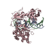
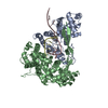
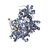
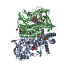
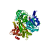
| Title | Structure of the class II 3-hydroxy-3-methylglutaryl-CoA reductase from Streptococcus pneumoniae bound to HMG-CoA and in a partially closed conformation | ||||||
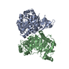
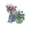
 Components Components | 3-hydroxy-3-methylglutaryl coenzyme A reductase | ||||||
 Keywords Keywords |  OXIDOREDUCTASE / 3-hydroxy-3-methylglutaryl-CoA reductase / HMG-CoA / conformational change OXIDOREDUCTASE / 3-hydroxy-3-methylglutaryl-CoA reductase / HMG-CoA / conformational change | ||||||
| Function / homology |  Function and homology information Function and homology informationhydroxymethylglutaryl-CoA reductase (NADH) activity / hydroxymethylglutaryl-CoA reductase / hydroxymethylglutaryl-CoA reductase (NADPH) activity / coenzyme A metabolic processSimilarity search - Function | ||||||
| Biological species |   Streptococcus pneumoniae (bacteria) Streptococcus pneumoniae (bacteria) | ||||||
| Method | X-RAY DIFFRACTION / SYNCHROTRON / MOLECULAR REPLACEMENT / Resolution: 2.3 Å | ||||||
 Authors Authors | Miller, B.R. / Kung, Y. | ||||||
| Funding support |  United States, 1items United States, 1items
| ||||||
 Citation Citation | Journal: Biochemistry / Year: 2018 Title: Structural Features and Domain Movements Controlling Substrate Binding and Cofactor Specificity in Class II HMG-CoA Reductase. Authors: Miller, B.R. / Kung, Y. | ||||||
| History |
|
- Structure visualization
| Structure viewer | Molecule: MolmilJmol/JSmol |
|---|
- Downloads & links
-Download
| PDBx/mmCIF format | 5wpk.cif.gz | 178.5 KB | Display | PDBx/mmCIF format |
|---|---|---|---|---|
| PDB format | pdb5wpk.ent.gz | 137.9 KB | Display | PDB format |
| PDBx/mmJSON format | 5wpk.json.gz | Tree view | PDBx/mmJSON format | |
| Others |  Other downloads Other downloads |
-Validation report
| Arichive directory | https://data.pdbj.org/pub/pdb/validation_reports/wp/5wpkftp://data.pdbj.org/pub/pdb/validation_reports/wp/5wpk | HTTPS FTP |
|---|
-Related structure data
| Related structure data |  5wpjC  3qaeS S: Starting model for refinement C: citing same article ( |
|---|---|
| Similar structure data |


-Links
 PDBj
PDBj
- Assembly
| Deposited unit | 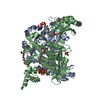
| ||||||||
|---|---|---|---|---|---|---|---|---|---|
| 1 |
| ||||||||
| Unit cell |
|
-Components
| #1: Protein | Mass: 46457.820 Da / Num. of mol.: 2 Source method: isolated from a genetically manipulated source Source: (gene. exp.) Streptococcus pneumoniae (bacteria)Gene: mvaA, mvaA_1, mvaA_3, AWW74_09880, ERS019416_00185, ERS019687_00346, ERS020135_00205, ERS020138_01692, ERS020141_01867, ERS020145_01986, ERS020146_01579, ERS020155_00051, ERS020158_01777, ...Gene: mvaA, mvaA_1, mvaA_3, AWW74_09880, ERS019416_00185, ERS019687_00346, ERS020135_00205, ERS020138_01692, ERS020141_01867, ERS020145_01986, ERS020146_01579, ERS020155_00051, ERS020158_01777, ERS020520_02006, ERS020525_01644, ERS020528_00536, ERS020531_01915, ERS020532_00514, ERS020539_01756, ERS020726_02213, ERS020726_02223, ERS020822_01277, ERS021354_03805, ERS021629_07183, ERS021733_05462, ERS232524_02161, ERS367337_00780, ERS409444_00519 Plasmid: pSKB3 Details (production host): N-terminal hexa-his-tag with kanomycin cassette Production host: Escherichia coli BL21(DE3) (bacteria)References: UniProt: A0A0D6J7E8, UniProt: Q8DNS5*PLUS, hydroxymethylglutaryl-CoA reductase (NADPH)#2: Chemical | HMG-CoA  Mass: 906.620 Da / Num. of mol.: 2 / Source method: obtained synthetically / Formula: C27H39N7O20P3S Mass: 906.620 Da / Num. of mol.: 2 / Source method: obtained synthetically / Formula: C27H39N7O20P3S#3: Chemical | Glycerol  Mass: 92.094 Da / Num. of mol.: 3 / Source method: obtained synthetically / Formula: C3H8O3 Mass: 92.094 Da / Num. of mol.: 3 / Source method: obtained synthetically / Formula: C3H8O3#4: Chemical | Polyethylene glycol  Mass: 354.436 Da / Num. of mol.: 2 / Source method: obtained synthetically / Formula: C16H34O8 / Comment: precipitant*YM Mass: 354.436 Da / Num. of mol.: 2 / Source method: obtained synthetically / Formula: C16H34O8 / Comment: precipitant*YM#5: Water | ChemComp-HOH / | Water Mass: 18.015 Da / Num. of mol.: 417 / Source method: isolated from a natural source / Formula: H2O Mass: 18.015 Da / Num. of mol.: 417 / Source method: isolated from a natural source / Formula: H2O |
|---|
-Experimental details
-Experiment
| Experiment | Method: X-RAY DIFFRACTION / Number of used crystals: 1 |
|---|
- Sample preparation
| Crystal | Density Matthews: 2.32 Å3/Da / Density % sol: 47.06 % |
|---|---|
| Crystal grow | Temperature: 295 K / Method: vapor diffusion, hanging drop / pH: 8.5 Details: 100 mM Tris, pH 8.5, 100-250 mM lithium sulfate, 15-25% PEG4000 Temp details: Room Temperature |
-Data collection
| Diffraction | Mean temperature: 90 K |
|---|---|
| Diffraction source | Source: SYNCHROTRON / Site: APS / Beamline: 24-ID-E / Wavelength: 0.9792 Å |
| Detector | Type: DECTRIS EIGER X 16M / Detector: PIXEL / Date: Jul 30, 2017 |
| Radiation | Monochromator: Si(220) / Protocol: SINGLE WAVELENGTH / Monochromatic (M) / Laue (L): M / Scattering type: x-ray |
| Radiation wavelength | Wavelength: 0.9792 Å / Relative weight: 1 |
| Reflection | Resolution: 2.3→19.241 Å / Num. obs: 36792 / % possible obs: 98.17 % / Redundancy: 3.5 % / CC1/2: 0.987 / Rmerge(I) obs: 0.1514 / Net I/σ(I): 6.48 |
| Reflection shell | Resolution: 2.3→2.382 Å / Redundancy: 3.5 % / Rmerge(I) obs: 0.5832 / Mean I/σ(I) obs: 1.84 / Num. unique obs: 3643 / CC1/2: 0.676 / % possible all: 97.98 |
- Processing
| Software |
| ||||||||||||||||||||||||||||||||||||||||||||||||||||||||||||||||||||||||||||||||||||||||||||||||||
|---|---|---|---|---|---|---|---|---|---|---|---|---|---|---|---|---|---|---|---|---|---|---|---|---|---|---|---|---|---|---|---|---|---|---|---|---|---|---|---|---|---|---|---|---|---|---|---|---|---|---|---|---|---|---|---|---|---|---|---|---|---|---|---|---|---|---|---|---|---|---|---|---|---|---|---|---|---|---|---|---|---|---|---|---|---|---|---|---|---|---|---|---|---|---|---|---|---|---|---|
| Refinement | Method to determine structure: MOLECULAR REPLACEMENT Starting model: PDB entry 3QAE Resolution: 2.3→19.241 Å / Cross valid method: FREE R-VALUE / σ(F): 478.83 / Phase error: 27.84
| ||||||||||||||||||||||||||||||||||||||||||||||||||||||||||||||||||||||||||||||||||||||||||||||||||
| Solvent computation | Shrinkage radii: 0.9 Å / VDW probe radii: 1.11 Å | ||||||||||||||||||||||||||||||||||||||||||||||||||||||||||||||||||||||||||||||||||||||||||||||||||
| Refinement step | Cycle: LAST / Resolution: 2.3→19.241 Å
| ||||||||||||||||||||||||||||||||||||||||||||||||||||||||||||||||||||||||||||||||||||||||||||||||||
| Refine LS restraints |
| ||||||||||||||||||||||||||||||||||||||||||||||||||||||||||||||||||||||||||||||||||||||||||||||||||
| LS refinement shell |
|
