 Movie
Movie Controller

[English] 日本語
 Yorodumi
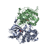
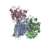
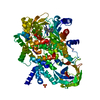
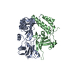
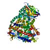
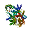
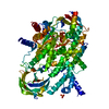
- PDB-3mbc: Crystal structure of monomeric isocitrate dehydrogenase from Cory... -
+ Open data

- Basic information
| Entry | Database: PDB / ID: 3mbc | ||||||
|---|---|---|---|---|---|---|---|
| Title | Crystal structure of monomeric isocitrate dehydrogenase from Corynebacterium glutamicum in complex with NADP | ||||||
 Components Components | Isocitrate dehydrogenase [NADP] | ||||||
 Keywords Keywords |  OXIDOREDUCTASE / Holoenzyme / Apoenzyme / Open conformation / NADP / Glyoxylate bypass / Magnesium / Metal-binding / Tricarboxylic acid cycle OXIDOREDUCTASE / Holoenzyme / Apoenzyme / Open conformation / NADP / Glyoxylate bypass / Magnesium / Metal-binding / Tricarboxylic acid cycle | ||||||
| Function / homology | Isocitrate dehydrogenase NADP-dependent, monomeric / Monomeric isocitrate dehydrogenase / isocitrate dehydrogenase (NADP+) / isocitrate dehydrogenase (NADP+) activity / glyoxylate cycle / tricarboxylic acid cycle / metal ion binding / NADP NICOTINAMIDE-ADENINE-DINUCLEOTIDE PHOSPHATE / Isocitrate dehydrogenase [NADP] Function and homology information Function and homology information | ||||||
| Biological species |  Corynebacterium glutamicum (bacteria) Corynebacterium glutamicum (bacteria) | ||||||
| Method | X-RAY DIFFRACTION / SYNCHROTRON / MOLECULAR REPLACEMENT / molecular replacement / Resolution: 1.9 Å | ||||||
 Authors Authors | Sidhu, N.S. / Aich, S. / Sheldrick, G.M. / Delbaere, L.T.J. | ||||||
 Citation Citation | Journal: Acta Crystallogr.,Sect.D / Year: 2011 Title: Structure of a highly NADP+-specific isocitrate dehydrogenase. Authors: Sidhu, N.S. / Delbaere, L.T. / Sheldrick, G.M. #1: Journal: Proteins / Year: 2006Title: Substrate-free structure of a monomeric NADP isocitrate dehydrogenase: an open conformation phylogenetic relationship of isocitrate dehydrogenase. Authors: Imabayashi, F. / Aich, S. / Prasad, L. / Delbaere, L.T. #2: Journal: Structure / Year: 2002Title: Structure of the monomeric isocitrate dehydrogenase: evidence of a protein monomerization by a domain duplication. Authors: Yasutake, Y. / Watanabe, S. / Yao, M. / Takada, Y. / Fukunaga, N. / Tanaka, I. #3: Journal: J.Biol.Chem. / Year: 2003Title: Crystal structure of the monomeric isocitrate dehydrogenase in the presence of NADP+: insight into the cofactor recognition, catalysis, and evolution. Authors: Yasutake, Y. / Watanabe, S. / Yao, M. / Takada, Y. / Fukunaga, N. / Tanaka, I. | ||||||
| History |
|
- Structure visualization
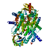
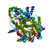
| Structure viewer | Molecule: MolmilJmol/JSmol |
|---|
- Downloads & links
-Download
| PDBx/mmCIF format | 3mbc.cif.gz | 595.9 KB | Display | PDBx/mmCIF format |
|---|---|---|---|---|
| PDB format | pdb3mbc.ent.gz | 492.5 KB | Display | PDB format |
| PDBx/mmJSON format | 3mbc.json.gz | Tree view | PDBx/mmJSON format | |
| Others |  Other downloads Other downloads |
-Validation report
| Arichive directory | https://data.pdbj.org/pub/pdb/validation_reports/mb/3mbcftp://data.pdbj.org/pub/pdb/validation_reports/mb/3mbc | HTTPS FTP |
|---|
-Related structure data
| Related structure data |  2b0tS S: Starting model for refinement |
|---|---|
| Similar structure data |


-Links
 PDBj
PDBj
- Assembly
| Deposited unit | 
| ||||||||
|---|---|---|---|---|---|---|---|---|---|
| 1 | 
| ||||||||
| 2 | 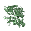
| ||||||||
| Unit cell |
|
-Components
| #1: Protein | Mass: 80167.008 Da / Num. of mol.: 2 / Source method: isolated from a natural source / Source: (natural) Corynebacterium glutamicum (bacteria) / Strain: ATCC 13032References: UniProt: P50216, isocitrate dehydrogenase (NADP+)#2: Chemical |   Mass: 24.305 Da / Num. of mol.: 2 / Source method: obtained synthetically / Formula: Mg Mass: 24.305 Da / Num. of mol.: 2 / Source method: obtained synthetically / Formula: Mg#3: Chemical | ChemComp-NAP / | Nicotinamide adenine dinucleotide phosphate  Mass: 743.405 Da / Num. of mol.: 1 / Source method: obtained synthetically / Formula: C21H28N7O17P3 Mass: 743.405 Da / Num. of mol.: 1 / Source method: obtained synthetically / Formula: C21H28N7O17P3#4: Water | ChemComp-HOH / | Water Mass: 18.015 Da / Num. of mol.: 796 / Source method: isolated from a natural source / Formula: H2O Mass: 18.015 Da / Num. of mol.: 796 / Source method: isolated from a natural source / Formula: H2O |
|---|
-Experimental details
-Experiment
| Experiment | Method: X-RAY DIFFRACTION / Number of used crystals: 1 |
|---|
- Sample preparation
| Crystal | Density Matthews: 2.44 Å3/Da / Density % sol: 49.5 % |
|---|---|
| Crystal grow | Temperature: 298 K / Method: vapor diffusion, hanging drop / pH: 7.3 Details: 25% polyethylene glycol 2000 monomethyl ether, 0.2 M tris[hydroxymethyl]aminomethane-HCl buffer, 0.2 M MgCl2, and 10 mM NADP, pH 7.3, VAPOR DIFFUSION, HANGING DROP, temperature 298K |
-Data collection
| Diffraction | Mean temperature: 100 K |
|---|---|
| Diffraction source | Source: SYNCHROTRON / Site: CLSI  / Beamline: 08ID-1 / Wavelength: 0.97934 Å / Beamline: 08ID-1 / Wavelength: 0.97934 Å |
| Detector | Type: MARMOSAIC 225 mm CCD / Detector: CCD / Date: Mar 18, 2008 |
| Radiation | Monochromator: Double crystal monochromator with indirectly cryo-cooled and sagittally focusing crystals Protocol: SINGLE WAVELENGTH / Monochromatic (M) / Laue (L): M / Scattering type: x-ray |
| Radiation wavelength | Wavelength: 0.97934 Å / Relative weight: 1 |
| Reflection | Resolution: 1.9→19.74 Å / Num. all: 122536 / Num. obs: 119827 / % possible obs: 97.8 % / Observed criterion σ(I): -3 / Redundancy: 3.66 % / Rmerge(I) obs: 0.088 / Net I/σ(I): 9.94 |
| Reflection shell | Resolution: 1.9→1.95 Å / Redundancy: 3.25 % / Rmerge(I) obs: 0.308 / Mean I/σ(I) obs: 3.5 / % possible all: 85.4 |
-Phasing
| Phasing | Method: molecular replacement | |||||||||
|---|---|---|---|---|---|---|---|---|---|---|
| Phasing MR | Model details: Phaser MODE: MR_AUTO
|
- Processing
| Software |
| |||||||||||||||||||||||||||||||||||||||||||||||||||||||||||||||||||||||||||||||||||||||||||||||||||||||||||||||||||||||||||||||||||||||||||||||||||||||||||||||||||||||||||||||
|---|---|---|---|---|---|---|---|---|---|---|---|---|---|---|---|---|---|---|---|---|---|---|---|---|---|---|---|---|---|---|---|---|---|---|---|---|---|---|---|---|---|---|---|---|---|---|---|---|---|---|---|---|---|---|---|---|---|---|---|---|---|---|---|---|---|---|---|---|---|---|---|---|---|---|---|---|---|---|---|---|---|---|---|---|---|---|---|---|---|---|---|---|---|---|---|---|---|---|---|---|---|---|---|---|---|---|---|---|---|---|---|---|---|---|---|---|---|---|---|---|---|---|---|---|---|---|---|---|---|---|---|---|---|---|---|---|---|---|---|---|---|---|---|---|---|---|---|---|---|---|---|---|---|---|---|---|---|---|---|---|---|---|---|---|---|---|---|---|---|---|---|---|---|---|---|---|
| Refinement | Method to determine structure: MOLECULAR REPLACEMENT Starting model: PDB entry 2b0t split into 2 domains: Domain I (residues 2-138, and 558-737) and Domain II (139-557). Resolution: 1.9→19.74 Å / Cor.coef. Fo:Fc: 0.944 / Cor.coef. Fo:Fc free: 0.92 / Occupancy max: 1 / Occupancy min: 0.5 / SU B: 7.725 / SU ML: 0.105 / SU R Cruickshank DPI: 0.157 / Isotropic thermal model: Isotropic / Cross valid method: THROUGHOUT / ESU R: 0.157 / ESU R Free: 0.144 / Stereochemistry target values: MAXIMUM LIKELIHOOD / Details: HYDROGENS HAVE BEEN ADDED IN THE RIDING POSITIONS
| |||||||||||||||||||||||||||||||||||||||||||||||||||||||||||||||||||||||||||||||||||||||||||||||||||||||||||||||||||||||||||||||||||||||||||||||||||||||||||||||||||||||||||||||
| Solvent computation | Ion probe radii: 0.8 Å / Shrinkage radii: 0.8 Å / VDW probe radii: 1.2 Å / Solvent model: MASK | |||||||||||||||||||||||||||||||||||||||||||||||||||||||||||||||||||||||||||||||||||||||||||||||||||||||||||||||||||||||||||||||||||||||||||||||||||||||||||||||||||||||||||||||
| Displacement parameters | Biso mean: 30.086 Å2
| |||||||||||||||||||||||||||||||||||||||||||||||||||||||||||||||||||||||||||||||||||||||||||||||||||||||||||||||||||||||||||||||||||||||||||||||||||||||||||||||||||||||||||||||
| Refine analyze | Luzzati coordinate error obs: 0.26 Å | |||||||||||||||||||||||||||||||||||||||||||||||||||||||||||||||||||||||||||||||||||||||||||||||||||||||||||||||||||||||||||||||||||||||||||||||||||||||||||||||||||||||||||||||
| Refinement step | Cycle: LAST / Resolution: 1.9→19.74 Å
| |||||||||||||||||||||||||||||||||||||||||||||||||||||||||||||||||||||||||||||||||||||||||||||||||||||||||||||||||||||||||||||||||||||||||||||||||||||||||||||||||||||||||||||||
| Refine LS restraints |
| |||||||||||||||||||||||||||||||||||||||||||||||||||||||||||||||||||||||||||||||||||||||||||||||||||||||||||||||||||||||||||||||||||||||||||||||||||||||||||||||||||||||||||||||
| LS refinement shell | Resolution: 1.9→1.949 Å / Total num. of bins used: 20
| |||||||||||||||||||||||||||||||||||||||||||||||||||||||||||||||||||||||||||||||||||||||||||||||||||||||||||||||||||||||||||||||||||||||||||||||||||||||||||||||||||||||||||||||
| Refinement TLS params. | Method: refined / Refine-ID: X-RAY DIFFRACTION
| |||||||||||||||||||||||||||||||||||||||||||||||||||||||||||||||||||||||||||||||||||||||||||||||||||||||||||||||||||||||||||||||||||||||||||||||||||||||||||||||||||||||||||||||
| Refinement TLS group |
|
