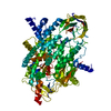
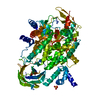
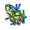
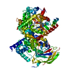
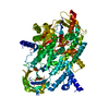
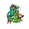
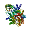
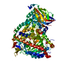
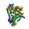
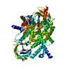
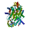
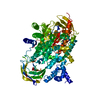
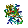
[English] 日本語
Yorodumi
- PDB-3qk0: Crystal structure of PI3K-gamma in complex with benzothiazole 82 -

+
Open data


ID or keywords:

Loading...

-
Basic information

Entry
Database: PDB / ID: 3qk0
TitleCrystal structure of PI3K-gamma in complex with benzothiazole 82
ComponentsPhosphatidylinositol-4,5-bisphosphate 3-kinase catalytic subunit gamma isoform
KeywordsTRANSFERASE/TRANSFERASE INHIBITOR / p110 / transferase / kinase / inhibitor / ATP-binding / p84 / p101 / TRANSFERASE-TRANSFERASE INHIBITOR complex
Function / homology
Function and homology information


secretory granule localization / negative regulation of triglyceride catabolic process / natural killer cell chemotaxis / neutrophil extravasation / phosphatidylinositol-4-phosphate 3-kinase / positive regulation of acute inflammatory response / respiratory burst involved in defense response / negative regulation of cardiac muscle contraction / regulation of calcium ion transmembrane transport / T cell chemotaxis ...secretory granule localization / negative regulation of triglyceride catabolic process / natural killer cell chemotaxis / neutrophil extravasation / phosphatidylinositol-4-phosphate 3-kinase / positive regulation of acute inflammatory response / respiratory burst involved in defense response / negative regulation of cardiac muscle contraction / regulation of calcium ion transmembrane transport / T cell chemotaxis / negative regulation of fibroblast apoptotic process / phosphatidylinositol 3-kinase complex, class IB / sphingosine-1-phosphate receptor signaling pathway / phosphatidylinositol 3-kinase complex, class IA / dendritic cell chemotaxis / 1-phosphatidylinositol-4-phosphate 3-kinase activity / 1-phosphatidylinositol-4,5-bisphosphate 3-kinase activity / phosphatidylinositol-4,5-bisphosphate 3-kinase / phosphatidylinositol 3-kinase / phosphatidylinositol-3-phosphate biosynthetic process / 1-phosphatidylinositol-3-kinase activity / mast cell degranulation / Erythropoietin activates Phosphoinositide-3-kinase (PI3K) / hepatocyte apoptotic process / positive regulation of Rac protein signal transduction / phosphatidylinositol phosphate biosynthetic process / phosphatidylinositol-mediated signaling / regulation of cell adhesion mediated by integrin / Synthesis of PIPs at the plasma membrane / regulation of angiogenesis / T cell proliferation / cellular response to cAMP / GPVI-mediated activation cascade / T cell activation / ephrin receptor binding / positive regulation of endothelial cell migration / neutrophil chemotaxis / phosphatidylinositol 3-kinase/protein kinase B signal transduction / positive regulation of cytokine production / positive regulation of MAP kinase activity / platelet aggregation / endocytosis / G beta:gamma signalling through PI3Kgamma / Signaling by CSF1 (M-CSF) in myeloid cells / kinase activity / positive regulation of cytosolic calcium ion concentration / angiogenesis / adaptive immune response / positive regulation of phosphatidylinositol 3-kinase/protein kinase B signal transduction / non-specific serine/threonine protein kinase / protein kinase activity / inflammatory response / immune response / G protein-coupled receptor signaling pathway / phosphorylation / protein serine kinase activity / innate immune response / protein serine/threonine kinase activity / ATP binding / membrane / identical protein binding / plasma membrane / cytosol / cytoplasm
Similarity search - Function
PIK3 catalytic subunit gamma, adaptor-binding domain / PIK3 catalytic subunit gamma adaptor-binding domain / Phosphatidylinositol 3-/4-kinase, catalytic domain / Phosphatidylinositol 3-kinase, accessory domain (PIK) / Phosphatidylinositol 3-kinase Catalytic Subunit; Chain A, domain 4 / Phosphatidylinositol 3-kinase Catalytic Subunit; Chain A, domain 4 / Phosphatidylinositol 3-kinase Catalytic Subunit; Chain A, Domain 5 / C2 domain / Phosphatidylinositol 3-kinase, adaptor-binding domain / Phosphatidylinositol 3-kinase adaptor-binding (PI3K ABD) domain profile. ...PIK3 catalytic subunit gamma, adaptor-binding domain / PIK3 catalytic subunit gamma adaptor-binding domain / Phosphatidylinositol 3-/4-kinase, catalytic domain / Phosphatidylinositol 3-kinase, accessory domain (PIK) / Phosphatidylinositol 3-kinase Catalytic Subunit; Chain A, domain 4 / Phosphatidylinositol 3-kinase Catalytic Subunit; Chain A, domain 4 / Phosphatidylinositol 3-kinase Catalytic Subunit; Chain A, Domain 5 / C2 domain / Phosphatidylinositol 3-kinase, adaptor-binding domain / Phosphatidylinositol 3-kinase adaptor-binding (PI3K ABD) domain profile. / PI3-kinase family, Ras-binding domain / Phosphatidylinositol 3-kinase Ras-binding (PI3K RBD) domain / PI3-kinase family, ras-binding domain / Phosphatidylinositol 3-kinase Ras-binding (PI3K RBD) domain profile. / Phosphoinositide 3-kinase C2 / Phosphoinositide 3-kinase, region postulated to contain C2 domain / C2 phosphatidylinositol 3-kinase-type domain / C2 phosphatidylinositol 3-kinase (PI3K)-type domain profile. / Phosphoinositide 3-kinase, accessory (PIK) domain superfamily / Phosphoinositide 3-kinase family, accessory domain (PIK domain) / Phosphoinositide 3-kinase family, accessory domain (PIK domain) / Phosphoinositide 3-kinase, accessory (PIK) domain / Phosphatidylinositol kinase / PIK helical domain profile. / Phosphatidylinositol 3- and 4-kinases signature 1. / Phosphatidylinositol 3/4-kinase, conserved site / Phosphatidylinositol 3- and 4-kinases signature 2. / Phosphatidylinositol 3-/4-kinase, catalytic domain superfamily / Phosphoinositide 3-kinase, catalytic domain / Phosphatidylinositol 3- and 4-kinase / Phosphatidylinositol 3- and 4-kinases catalytic domain profile. / Phosphatidylinositol 3-/4-kinase, catalytic domain / Phosphatidylinositol 3-kinase Catalytic Subunit; Chain A, domain 1 / C2 domain superfamily / Serine Threonine Protein Phosphatase 5, Tetratricopeptide repeat / Ubiquitin-like (UB roll) / Alpha Horseshoe / Armadillo-type fold / Ubiquitin-like domain superfamily / Roll / Protein kinase-like domain superfamily / Immunoglobulin-like / Sandwich / 2-Layer Sandwich / Orthogonal Bundle / Mainly Beta / Mainly Alpha / Alpha Beta
Similarity search - Domain/homology
Chem-QK0 / Phosphatidylinositol 4,5-bisphosphate 3-kinase catalytic subunit gamma isoform
Similarity search - Component
Biological speciesHomo sapiens (human)
MethodX-RAY DIFFRACTION / MOLECULAR REPLACEMENT / Resolution: 2.85 Å
AuthorsWhittington, D.A. / Tang, J. / Yakowec, P.
CitationJournal: J.Med.Chem. / Year: 2011
Title: Discovery and Optimization of a Series of Benzothiazole Phosphoinositide 3-Kinase (PI3K)/Mammalian Target of Rapamycin (mTOR) Dual Inhibitors.
Authors: D'Angelo, N.D. / Kim, T.S. / Andrews, K. / Booker, S.K. / Caenepeel, S. / Chen, K. / D'Amico, D. / Freeman, D. / Jiang, J. / Liu, L. / McCarter, J.D. / San Miguel, T. / Mullady, E.L. / ...Authors: D'Angelo, N.D. / Kim, T.S. / Andrews, K. / Booker, S.K. / Caenepeel, S. / Chen, K. / D'Amico, D. / Freeman, D. / Jiang, J. / Liu, L. / McCarter, J.D. / San Miguel, T. / Mullady, E.L. / Schrag, M. / Subramanian, R. / Tang, J. / Wahl, R.C. / Wang, L. / Whittington, D.A. / Wu, T. / Xi, N. / Xu, Y. / Yakowec, P. / Yang, K. / Zalameda, L.P. / Zhang, N. / Hughes, P. / Norman, M.H.
History
DepositionJan 31, 2011Deposition site: RCSB / Processing site: RCSB
Revision 1.0Mar 30, 2011Provider: repository / Type: Initial release
Revision 1.1Jul 13, 2011Group: Version format compliance
Revision 1.2Sep 13, 2023Group: Data collection / Database references ...Data collection / Database references / Derived calculations / Refinement description
Category: chem_comp_atom / chem_comp_bond ...chem_comp_atom / chem_comp_bond / database_2 / pdbx_initial_refinement_model / struct_ref_seq_dif / struct_site
Item: _database_2.pdbx_DOI / _database_2.pdbx_database_accession ..._database_2.pdbx_DOI / _database_2.pdbx_database_accession / _struct_ref_seq_dif.details / _struct_site.pdbx_auth_asym_id / _struct_site.pdbx_auth_comp_id / _struct_site.pdbx_auth_seq_id

-
Structure visualization

Structure viewerMolecule:
MolmilJmol/JSmol

Downloads & links

-
Assembly

Deposited unit
A: Phosphatidylinositol-4,5-bisphosphate 3-kinase catalytic subunit gamma isoform
hetero molecules


Theoretical massNumber of molelcules
Total (without water)110,5895
Polymers109,8241
Non-polymers7654
Water1267
1


  • Idetical with deposited unit
  • defined by author
TypeNameSymmetry operationNumber
identity operation1_555x,y,z1
Unit cell
Length a, b, c (Å)144.525, 67.835, 107.096
Angle α, β, γ (deg.)90.00, 95.34, 90.00
Int Tables number5
Space group name H-MC121

-
Components

#1: Protein Phosphatidylinositol-4,5-bisphosphate 3-kinase catalytic subunit gamma isoform / PI3-kinase subunit gamma / PI3K-gamma / PtdIns-3-kinase subunit gamma / Phosphatidylinositol-4 / 5- ...PI3-kinase subunit gamma / PI3K-gamma / PtdIns-3-kinase subunit gamma / Phosphatidylinositol-4 / 5-bisphosphate 3-kinase 110 kDa catalytic subunit gamma / PtdIns-3-kinase subunit p110-gamma / p120-PI3K


Mass: 109824.055 Da / Num. of mol.: 1 / Fragment: catalytic domain (UNP Residues 144-1102)
Source method: isolated from a genetically manipulated source
Source: (gene. exp.) Homo sapiens (human) / Gene: PIK3CG / Production host: Spodoptera frugiperda (fall armyworm)
References: UniProt: P48736, phosphatidylinositol-4,5-bisphosphate 3-kinase
#2: Chemical ChemComp-SO4 / SULFATE ION / Sulfate


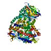
Mass: 96.063 Da / Num. of mol.: 3 / Source method: obtained synthetically / Formula: SO4
#3: Chemical ChemComp-QK0 / N-[6-(6-chloro-5-{[(4-fluorophenyl)sulfonyl]amino}pyridin-3-yl)-1,3-benzothiazol-2-yl]acetamide


Mass: 476.932 Da / Num. of mol.: 1 / Source method: obtained synthetically / Formula: C20H14ClFN4O3S2
#4: Water ChemComp-HOH / water / Water


Mass: 18.015 Da / Num. of mol.: 7 / Source method: isolated from a natural source / Formula: H2O

-
Experimental details

-
Experiment

ExperimentMethod: X-RAY DIFFRACTION / Number of used crystals: 1

-
Sample preparation

CrystalDensity Matthews: 2.38 Å3/Da / Density % sol: 48.31 %
Crystal growTemperature: 293 K / Method: vapor diffusion, hanging drop / pH: 7.5
Details: 21% PEG 3350, 240 mM ammonium sulfate, 2 mM dithiothreitol, 100 mM Tris, pH 7.5, VAPOR DIFFUSION, HANGING DROP, temperature 293K

-
Data collection

DiffractionMean temperature: 100 K
Diffraction sourceSource: ROTATING ANODE / Type: RIGAKU FR-E SUPERBRIGHT / Wavelength: 1.5418 Å
DetectorType: RIGAKU RAXIS IV++ / Detector: IMAGE PLATE / Date: Sep 10, 2007
RadiationProtocol: SINGLE WAVELENGTH / Monochromatic (M) / Laue (L): M / Scattering type: x-ray
Radiation wavelengthWavelength: 1.5418 Å / Relative weight: 1
ReflectionResolution: 2.85→30 Å / Num. all: 24355 / Num. obs: 24209 / % possible obs: 99.4 % / Observed criterion σ(I): -3 / Redundancy: 3.4 % / Biso Wilson estimate: 96.2 Å2 / Rmerge(I) obs: 0.058 / Χ2: 1.183 / Net I/σ(I): 18.9
Reflection shell
Resolution (Å)Redundancy (%)Rmerge(I) obsMean I/σ(I) obsNum. unique allΧ2% possible all
2.85-2.9530.5742.1923381.52296.9
2.95-3.073.30.45923951.51499.2
3.07-3.213.40.33324041.46599.9
3.21-3.383.40.21824141.4499.9
3.38-3.593.50.14124041.43499.8
3.59-3.873.50.08824201.28399.9
3.87-4.253.50.05724470.987100
4.25-4.873.50.04124470.7899.9
4.87-6.123.50.03624510.66199.9
6.12-303.40.02824890.88199.1

-
Processing

Software
NameVersionClassificationNB
SCALEPACKdata scaling
REFMACrefinement
PDB_EXTRACT3.1data extraction
DENZOdata reduction
EPMRphasing
RefinementMethod to determine structure: MOLECULAR REPLACEMENT
Starting model: PDB ENTRY 1E8Y
Resolution: 2.85→30 Å / Cor.coef. Fo:Fc: 0.938 / Cor.coef. Fo:Fc free: 0.908 / WRfactor Rfree: 0.2957 / WRfactor Rwork: 0.2409 / Occupancy max: 1 / Occupancy min: 1 / FOM work R set: 0.7752 / SU B: 44.307 / SU ML: 0.387 / SU R Cruickshank DPI: 0.3697 / SU Rfree: 0.4484 / Cross valid method: THROUGHOUT / σ(F): 0 / ESU R Free: 0.438 / Stereochemistry target values: Engh & Huber / Details: HYDROGENS HAVE BEEN ADDED IN THE RIDING POSITIONS
RfactorNum. reflection% reflectionSelection details
Rfree0.2771 1432 5.9 %RANDOM
Rwork0.2258 ---
obs0.2289 24207 99.44 %-
Solvent computationIon probe radii: 0.8 Å / Shrinkage radii: 0.8 Å / VDW probe radii: 1.2 Å / Solvent model: MASK
Displacement parametersBiso max: 168.56 Å2 / Biso mean: 90.0258 Å2 / Biso min: 42.03 Å2
Baniso -1Baniso -2Baniso -3
1--1.83 Å20 Å20.88 Å2
2--2.5 Å20 Å2
3----0.51 Å2
Refinement stepCycle: LAST / Resolution: 2.85→30 Å
ProteinNucleic acidLigandSolventTotal
Num. atoms6638 0 46 7 6691
Refine LS restraints
Refine-IDTypeDev idealDev ideal targetNumber
X-RAY DIFFRACTIONr_bond_refined_d0.0070.0226824
X-RAY DIFFRACTIONr_angle_refined_deg1.0541.9669237
X-RAY DIFFRACTIONr_dihedral_angle_1_deg5.3295811
X-RAY DIFFRACTIONr_dihedral_angle_2_deg36.89424.272316
X-RAY DIFFRACTIONr_dihedral_angle_3_deg16.582151235
X-RAY DIFFRACTIONr_dihedral_angle_4_deg13.0121540
X-RAY DIFFRACTIONr_chiral_restr0.0720.21040
X-RAY DIFFRACTIONr_gen_planes_refined0.0030.025059
X-RAY DIFFRACTIONr_nbd_refined0.1990.23135
X-RAY DIFFRACTIONr_nbtor_refined0.30.24664
X-RAY DIFFRACTIONr_xyhbond_nbd_refined0.1350.2195
X-RAY DIFFRACTIONr_symmetry_vdw_refined0.2230.234
X-RAY DIFFRACTIONr_symmetry_hbond_refined0.1650.21
X-RAY DIFFRACTIONr_mcbond_it0.3691.54198
X-RAY DIFFRACTIONr_mcangle_it0.66726632
X-RAY DIFFRACTIONr_scbond_it0.65632983
X-RAY DIFFRACTIONr_scangle_it1.0844.52605
LS refinement shellResolution: 2.85→2.923 Å / Total num. of bins used: 20
RfactorNum. reflection% reflection
Rfree0.42 91 -
Rwork0.362 1589 -
all-1680 -
obs--96.28 %
Refinement TLS params.

Method: refined / Refine-ID: X-RAY DIFFRACTION

IDL112)L122)L132)L222)L232)L332)S11 (Å °)S12 (Å °)S13 (Å °)S21 (Å °)S22 (Å °)S23 (Å °)S31 (Å °)S32 (Å °)S33 (Å °)T112)T122)T132)T222)T232)T332)Origin x (Å)Origin y (Å)Origin z (Å)
15.3072-0.56310.38760.9924-0.58562.76480.1103-0.0926-1.2413-0.0130.27770.57310.2522-0.514-0.388-0.2431-0.0601-0.0035-0.31990.2080.188421.6977-13.783728.465
24.9895-1.8368-0.30495.19222.3188.02390.4684-0.6224-0.93340.09650.37040.841-0.0662-1.4917-0.8388-0.4439-0.05260.09550.06950.48020.319412.0723-11.885731.9709
32.72961.1068-1.40133.6894-1.6144.98830.02370.26640.1252-0.06190.12060.0429-0.10180.8051-0.1443-0.2118-0.02830.03850.3804-0.113-0.237566.7294-4.267414.3749
44.2511-1.9272-0.17633.8602-1.86913.81880.00310.7379-0.02870.0456-0.0071-0.061-0.11430.61940.004-0.1591-0.00770.01090.3138-0.1681-0.196756.617-6.069312.3122
53.6498-0.52862.12780.243-0.17982.52260.1074-0.0694-0.50160.07970.11790.1762-0.01830.1076-0.2253-0.02640.01410.0341-0.13560.01390.019946.1521-10.019534.4142
63.8326-1.17331.57821.7544-0.54742.62240.07770.5096-0.10130.07620.11040.2428-0.4417-0.1874-0.1881-0.07060.11160.0656-0.02570.0925-0.044720.16765.078217.462
74.9392-1.36822.11452.2002-0.34261.584-0.8924-0.39510.84390.54560.2898-0.1874-0.6608-0.15180.60250.39870.2058-0.0963-0.2124-0.1031-0.047529.73519.738537.1361
Refinement TLS group
IDRefine-IDRefine TLS-IDAuth asym-IDAuth seq-ID
1X-RAY DIFFRACTION1A147 - 248
2X-RAY DIFFRACTION2A269 - 320
3X-RAY DIFFRACTION3A357 - 436
4X-RAY DIFFRACTION4A457 - 522
5X-RAY DIFFRACTION5A545 - 725
6X-RAY DIFFRACTION6A726 - 885
7X-RAY DIFFRACTION7A886 - 1092

+
About Yorodumi

-
News

-
Feb 9, 2022. New format data for meta-information of EMDB entries

New format data for meta-information of EMDB entries

  • Version 3 of the EMDB header file is now the official format.
  • The previous official version 1.9 will be removed from the archive.

Related info.:EMDB header

External links:wwPDB to switch to version 3 of the EMDB data model

-
Aug 12, 2020. Covid-19 info

Covid-19 info

URL: https://pdbj.org/emnavi/covid19.php

New page: Covid-19 featured information page in EM Navigator.

Related info.:Covid-19 info / Mar 5, 2020. Novel coronavirus structure data

+
Mar 5, 2020. Novel coronavirus structure data

Novel coronavirus structure data

Related info.:Yorodumi Speices / Aug 12, 2020. Covid-19 info

External links:COVID-19 featured content - PDBj / Molecule of the Month (242):Coronavirus Proteases

+
Jan 31, 2019. EMDB accession codes are about to change! (news from PDBe EMDB page)

EMDB accession codes are about to change! (news from PDBe EMDB page)

  • The allocation of 4 digits for EMDB accession codes will soon come to an end. Whilst these codes will remain in use, new EMDB accession codes will include an additional digit and will expand incrementally as the available range of codes is exhausted. The current 4-digit format prefixed with “EMD-” (i.e. EMD-XXXX) will advance to a 5-digit format (i.e. EMD-XXXXX), and so on. It is currently estimated that the 4-digit codes will be depleted around Spring 2019, at which point the 5-digit format will come into force.
  • The EM Navigator/Yorodumi systems omit the EMD- prefix.

Related info.:Q: What is EMD? / ID/Accession-code notation in Yorodumi/EM Navigator

External links:EMDB Accession Codes are Changing Soon! / Contact to PDBj

+
Jul 12, 2017. Major update of PDB

Major update of PDB

  • wwPDB released updated PDB data conforming to the new PDBx/mmCIF dictionary.
  • This is a major update changing the version number from 4 to 5, and with Remediation, in which all the entries are updated.
  • In this update, many items about electron microscopy experimental information are reorganized (e.g. em_software).
  • Now, EM Navigator and Yorodumi are based on the updated data.

External links:wwPDB Remediation / Enriched Model Files Conforming to OneDep Data Standards Now Available in the PDB FTP Archive

-
Yorodumi

Thousand views of thousand structures

  • Yorodumi is a browser for structure data from EMDB, PDB, SASBDB, etc.
  • This page is also the successor to EM Navigator detail page, and also detail information page/front-end page for Omokage search.
  • The word "yorodu" (or yorozu) is an old Japanese word meaning "ten thousand". "mi" (miru) is to see.

Related info.:EMDB / PDB / SASBDB / Comparison of 3 databanks / Yorodumi Search / Aug 31, 2016. New EM Navigator & Yorodumi / Yorodumi Papers / Jmol/JSmol / Function and homology information / Changes in new EM Navigator and Yorodumi

Read more