 Movie
Movie Controller


+ Open data

- Basic information
| Entry | Database: PDB / ID: 6iat | |||||||||||||||
|---|---|---|---|---|---|---|---|---|---|---|---|---|---|---|---|---|
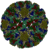
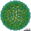
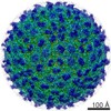
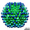
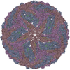
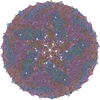
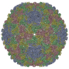
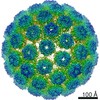
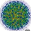
| Title | Icosahedrally averaged capsid of bacteriophage P68 | |||||||||||||||
 Components Components |
| |||||||||||||||
 Keywords Keywords |  STRUCTURAL PROTEIN / bacteriophage capsid / HK97 fold STRUCTURAL PROTEIN / bacteriophage capsid / HK97 fold | |||||||||||||||
| Function / homology | Uncharacterized protein / Major head protein Function and homology information Function and homology information | |||||||||||||||
| Biological species |  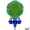 Staphylococcus phage P68 (virus) Staphylococcus phage P68 (virus) | |||||||||||||||
| Method | ELECTRON MICROSCOPY / single particle reconstruction / cryo EM / Resolution: 3.3 Å | |||||||||||||||
 Authors Authors | Hrebik, D. / Skubnik, K. / Fuzik, T. / Plevka, P. | |||||||||||||||
| Funding support |  Czech Republic, 4items Czech Republic, 4items
| |||||||||||||||
 Citation Citation | Journal: Sci Adv / Year: 2019 Title: Structure and genome ejection mechanism of phage P68. Authors: Dominik Hrebík / Dana Štveráková / Karel Škubník / Tibor Füzik / Roman Pantůček / Pavel Plevka / Abstract: Phages infecting can be used as therapeutics against antibiotic-resistant bacterial infections. However, there is limited information about the mechanism of genome delivery of phages that infect ...Phages infecting can be used as therapeutics against antibiotic-resistant bacterial infections. However, there is limited information about the mechanism of genome delivery of phages that infect Gram-positive bacteria. Here, we present the structures of native phage P68, genome ejection intermediate, and empty particle. The P68 head contains 72 subunits of inner core protein, 15 of which bind to and alter the structure of adjacent major capsid proteins and thus specify attachment sites for head fibers. Unlike in the previously studied phages, the head fibers of P68 enable its virion to position itself at the cell surface for genome delivery. The unique interaction of one end of P68 DNA with one of the 12 portal protein subunits is disrupted before the genome ejection. The inner core proteins are released together with the DNA and enable the translocation of phage genome across the bacterial membrane into the cytoplasm. | |||||||||||||||
| History |
|
- Structure visualization
| Movie |
Movie viewer |
|---|---|
| Structure viewer | Molecule: MolmilJmol/JSmol |
- Downloads & links
-Download
| PDBx/mmCIF format | 6iat.cif.gz | 313.9 KB | Display | PDBx/mmCIF format |
|---|---|---|---|---|
| PDB format | pdb6iat.ent.gz | 257.3 KB | Display | PDB format |
| PDBx/mmJSON format | 6iat.json.gz | Tree view | PDBx/mmJSON format | |
| Others |  Other downloads Other downloads |
-Validation report
| Arichive directory | https://data.pdbj.org/pub/pdb/validation_reports/ia/6iatftp://data.pdbj.org/pub/pdb/validation_reports/ia/6iat | HTTPS FTP |
|---|
-Related structure data
| Related structure data |  4438MC  4435C 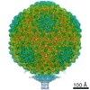 4436C  4437C  4440C 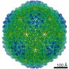 4442C  4449C  4450C 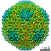 4451C  4453C  4454C  4455C 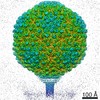 4456C  4457C  4458C 4459C  6iabC 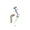 6iacC  6iawC  6ib1C  6q3gC M: map data used to model this data C: citing same article ( |
|---|---|
| Similar structure data |

-Links
 PDBj
PDBj- Assembly
| Deposited unit | 
|
|---|---|
| 1 | x 60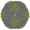
|
| 2 |
|
| 3 | x 5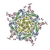
|
| 4 | x 6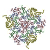
|
| 5 | 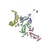
|
-Components
| #1: Protein | Mass: 46954.941 Da / Num. of mol.: 4 / Source method: isolated from a natural source / Source: (natural) Staphylococcus phage P68 (virus) / References: UniProt: Q859I3#2: Protein | Mass: 6922.464 Da / Num. of mol.: 4 / Source method: isolated from a natural source / Source: (natural) Staphylococcus phage P68 (virus) / References: UniProt: Q859I2 |
|---|
-Experimental details
-Experiment
| Experiment | Method: ELECTRON MICROSCOPY |
|---|---|
| EM experiment | Aggregation state: PARTICLE / 3D reconstruction method: single particle reconstruction |
- Sample preparation
| Component |
| ||||||||||||||||||||||||
|---|---|---|---|---|---|---|---|---|---|---|---|---|---|---|---|---|---|---|---|---|---|---|---|---|---|
| Molecular weight |
| ||||||||||||||||||||||||
| Source (natural) |
| ||||||||||||||||||||||||
| Details of virus | Empty: NO / Enveloped: NO / Isolate: STRAIN / Type: VIRION | ||||||||||||||||||||||||
| Natural host | Organism: Staphylococcus aureus / Strain: dTarM 4220 | ||||||||||||||||||||||||
| Virus shell | Name: Capsid / Diameter: 480 nm / Triangulation number (T number): 4 | ||||||||||||||||||||||||
| Buffer solution | pH: 8 | ||||||||||||||||||||||||
| Buffer component |
| ||||||||||||||||||||||||
| Specimen | Conc.: 2 mg/ml / Embedding applied: NO / Shadowing applied: NO / Staining applied: NO / Vitrification applied: YES | ||||||||||||||||||||||||
| Specimen support | Grid material: COPPER / Grid mesh size: 200 divisions/in. / Grid type: Quantifoil R2/1 | ||||||||||||||||||||||||
| Vitrification | Instrument: FEI VITROBOT MARK IV / Cryogen name: ETHANE / Humidity: 100 % / Chamber temperature: 293 K / Details: blot time 2s; blot force -2; 3.6 ul of sample |
- Electron microscopy imaging
| Experimental equipment |  Model: Titan Krios / Image courtesy: FEI Company |
|---|---|
| Microscopy | Model: FEI TITAN KRIOS |
| Electron gun | Electron source: FIELD EMISSION GUN / Accelerating voltage: 300 kV / Illumination mode: FLOOD BEAM |
| Electron lens | Mode: BRIGHT FIELDBright-field microscopy / Nominal magnification: 75000 X / Nominal defocus max: 3 nm / Nominal defocus min: 1 nm / Cs: 2.7 mm / C2 aperture diameter: 70 µm / Alignment procedure: COMA FREE |
| Specimen holder | Cryogen: NITROGEN / Specimen holder model: FEI TITAN KRIOS AUTOGRID HOLDER |
| Image recording | Average exposure time: 1 sec. / Electron dose: 21 e/Å2 / Detector mode: INTEGRATING / Film or detector model: FEI FALCON II (4k x 4k) / Num. of grids imaged: 2 / Num. of real images: 2891 |
| Image scans | Width: 4096 / Height: 4096 / Movie frames/image: 7 / Used frames/image: 1-7 |
- Processing
| Software | Name: PHENIX / Version: (dev_3042: ???) / Classification: refinement | ||||||||||||||||||||||||||||||||||||||||
|---|---|---|---|---|---|---|---|---|---|---|---|---|---|---|---|---|---|---|---|---|---|---|---|---|---|---|---|---|---|---|---|---|---|---|---|---|---|---|---|---|---|
| EM software |
| ||||||||||||||||||||||||||||||||||||||||
| CTF correction | Type: PHASE FLIPPING AND AMPLITUDE CORRECTION | ||||||||||||||||||||||||||||||||||||||||
| Particle selection | Num. of particles selected: 37218 / Details: Manual selection in EMAN | ||||||||||||||||||||||||||||||||||||||||
| Symmetry | Point symmetry: I (icosahedral) | ||||||||||||||||||||||||||||||||||||||||
| 3D reconstruction | Resolution: 3.3 Å / Resolution method: FSC 0.143 CUT-OFF / Num. of particles: 21625 / Num. of class averages: 1 / Symmetry type: POINT | ||||||||||||||||||||||||||||||||||||||||
| Atomic model building | Protocol: AB INITIO MODEL / Space: REAL | ||||||||||||||||||||||||||||||||||||||||
| Refinement | Resolution: 3.3→360.794 Å / SU ML: 0.67 / σ(F): 100 / Phase error: 34.78 / Stereochemistry target values: ML
| ||||||||||||||||||||||||||||||||||||||||
| Solvent computation | Shrinkage radii: 0.9 Å / VDW probe radii: 1.11 Å / Solvent model: FLAT BULK SOLVENT MODEL | ||||||||||||||||||||||||||||||||||||||||
| Refine LS restraints |
|
