[English] 日本語
Yorodumi
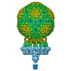
- EMDB-4459: Structure of native bacteriophage P68 -

+
Open data


ID or keywords:

Loading...

-
Basic information

Entry
Database: EMDB / ID: EMD-4459
TitleStructure of native bacteriophage P68
Map dataNone
Sample
  • Virus: Staphylococcus phage P68 (virus)
    • Complex: Capsid of native phage P68
      • Protein or peptide: Major head protein
      • Protein or peptide: Arstotzka protein
      • Protein or peptide: Head fiber protein
    • Complex: Tail complex
      • Protein or peptide: Lower collar protein
      • Protein or peptide: Minor structural protein
    • Complex: Portal protein
      • Protein or peptide: Portal protein
    • Complex: Inner core proteinEarth's inner core
      • Protein or peptide: Tail fibre protein
      • Protein or peptide: Inner core proteinEarth's inner core
Function / homology: / Phage 5-bladed beta propeller receptor binding platform domain / Uncharacterized protein / Uncharacterized protein / Major head protein / Lower collar protein / Minor structural protein
Function and homology information
Biological speciesStaphylococcus phage P68 (virus)
Methodsingle particle reconstruction / cryo EM / Resolution: 3.8 Å
AuthorsDominik H / Karel S / Fuzik T / Plevka P
Funding support Czech Republic, 3 items
OrganizationGrant numberCountry
Czech Science Foundation15-21631Y Czech Republic
European Molecular Biology Organization3041 Czech Republic
Czech Science Foundation18-17810S Czech Republic
CitationJournal: Sci Adv / Year: 2019
Title: Structure and genome ejection mechanism of phage P68.
Authors: Dominik Hrebík / Dana Štveráková / Karel Škubník / Tibor Füzik / Roman Pantůček / Pavel Plevka /
Abstract: Phages infecting can be used as therapeutics against antibiotic-resistant bacterial infections. However, there is limited information about the mechanism of genome delivery of phages that infect ...Phages infecting can be used as therapeutics against antibiotic-resistant bacterial infections. However, there is limited information about the mechanism of genome delivery of phages that infect Gram-positive bacteria. Here, we present the structures of native phage P68, genome ejection intermediate, and empty particle. The P68 head contains 72 subunits of inner core protein, 15 of which bind to and alter the structure of adjacent major capsid proteins and thus specify attachment sites for head fibers. Unlike in the previously studied phages, the head fibers of P68 enable its virion to position itself at the cell surface for genome delivery. The unique interaction of one end of P68 DNA with one of the 12 portal protein subunits is disrupted before the genome ejection. The inner core proteins are released together with the DNA and enable the translocation of phage genome across the bacterial membrane into the cytoplasm.
History
DepositionDec 4, 2018-
Header (metadata) releaseNov 6, 2019-
Map releaseNov 6, 2019-
UpdateNov 6, 2019-
Current statusNov 6, 2019Processing site: PDBe / Status: Released

-
Structure visualization

Movie
  • Surface view with section colored by density value
  • Surface level: 0.025
  • Imaged by UCSF Chimera
  • Download
  • Surface view colored by radius
  • Surface level: 0.025
  • Imaged by UCSF Chimera
  • Download
  • Surface view with fitted model
  • Atomic models: PDB-6q3g
  • Surface level: 0.025
  • Imaged by UCSF Chimera
  • Download
  • Simplified surface model + fitted atomic model
  • Atomic modelsPDB-6q3g
  • Imaged by Jmol
  • Download
Movie viewer
Structure viewerEM map:
SurfViewMolmilJmol/JSmol
Supplemental images

Downloads & links

-
Map

FileDownload / File: emd_4459.map.gz / Format: CCP4 / Size: 3.7 GB / Type: IMAGE STORED AS FLOATING POINT NUMBER (4 BYTES)
AnnotationNone
Voxel sizeX=Y=Z: 1.063 Å
Density
Contour LevelBy AUTHOR: 0.025 / Movie #1: 0.025
Minimum - Maximum-0.19396262 - 0.41469824
Average (Standard dev.)0.0007894909 (±0.009443255)
SymmetrySpace group: 1
Details

EMDB XML:

Map geometry
Axis orderXYZ
Origin-500-500-500
Dimensions100010001000
Spacing100010001000
CellA=B=C: 1063.0 Å
α=β=γ: 90.0 °

CCP4 map header:

modeImage stored as Reals
Å/pix. X/Y/Z1.0631.0631.063
M x/y/z100010001000
origin x/y/z0.0000.0000.000
length x/y/z1063.0001063.0001063.000
α/β/γ90.00090.00090.000
MAP C/R/S123
start NC/NR/NS-500-500-500
NC/NR/NS100010001000
D min/max/mean-0.1940.4150.001

-
Supplemental data

-
Additional map: Masked asymmetric reconstruction of native bacteriophage P68

Fileemd_4459_additional.map
AnnotationMasked asymmetric reconstruction of native bacteriophage P68
Projections & Slices
AxesZYX

Projections

Slices (1/2)
Density Histograms

-
Sample components

+
Entire : Staphylococcus phage P68

EntireName: Staphylococcus phage P68 (virus)
Components
  • Virus: Staphylococcus phage P68 (virus)
    • Complex: Capsid of native phage P68
      • Protein or peptide: Major head protein
      • Protein or peptide: Arstotzka protein
      • Protein or peptide: Head fiber protein
    • Complex: Tail complex
      • Protein or peptide: Lower collar protein
      • Protein or peptide: Minor structural protein
    • Complex: Portal protein
      • Protein or peptide: Portal protein
    • Complex: Inner core proteinEarth's inner core
      • Protein or peptide: Tail fibre protein
      • Protein or peptide: Inner core proteinEarth's inner core

+
Supramolecule #1: Staphylococcus phage P68

SupramoleculeName: Staphylococcus phage P68 / type: virus / ID: 1 / Parent: 0 / Macromolecule list: all / Virus type: VIRION / Virus isolate: STRAIN / Virus enveloped: No / Virus empty: No
Host (natural)Organism: Staphylococcus aureus (bacteria) / Strain: dTarM 4220
Molecular weightTheoretical: 19.7 MDa
Virus shellShell ID: 1 / Name: Capsid / Diameter: 480.0 Å / T number (triangulation number): 4

+
Supramolecule #2: Capsid of native phage P68

SupramoleculeName: Capsid of native phage P68 / type: complex / ID: 2 / Parent: 1 / Macromolecule list: #1-#2, #7
Source (natural)Organism: Staphylococcus phage P68 (virus)

+
Supramolecule #3: Tail complex

SupramoleculeName: Tail complex / type: complex / ID: 3 / Parent: 1 / Macromolecule list: #4-#5
Source (natural)Organism: Staphylococcus phage P68 (virus)

+
Supramolecule #4: Portal protein

SupramoleculeName: Portal protein / type: complex / ID: 4 / Parent: 1 / Macromolecule list: #3
Source (natural)Organism: Staphylococcus phage P68 (virus)

+
Supramolecule #5: Inner core protein

SupramoleculeName: Inner core protein / type: complex / ID: 5 / Parent: 1 / Macromolecule list: #6, #8
Source (natural)Organism: Staphylococcus phage P68 (virus)

+
Macromolecule #1: Major head protein

MacromoleculeName: Major head protein / type: protein_or_peptide / ID: 1 / Number of copies: 235 / Enantiomer: LEVO
Source (natural)Organism: Staphylococcus phage P68 (virus)
Molecular weightTheoretical: 46.954941 KDa
SequenceString: MAQQSTKNET ALLVAKSAKS ALQDFNHDYS KSWTFGDKWD NSNTMFETFV NKYLFPKINE TLLIDIALGN RFNWLAKEQD FIGQYSEEY VIMDTVPINM DLSKNEELML KRNYPRMATK LYGNGIVKKQ KFTLNNNDTR FNFQTLADAT NYALGVYKKK I SDINVLEE ...String:
MAQQSTKNET ALLVAKSAKS ALQDFNHDYS KSWTFGDKWD NSNTMFETFV NKYLFPKINE TLLIDIALGN RFNWLAKEQD FIGQYSEEY VIMDTVPINM DLSKNEELML KRNYPRMATK LYGNGIVKKQ KFTLNNNDTR FNFQTLADAT NYALGVYKKK I SDINVLEE KEMRAMLVDY SLNQLSETNV RKATSKEDLA SKVFEAILNL QNNSAKYNEV HRASGGAIGQ YTTVSKLKDI VI LTTDSLK SYLLDTKIAN TFQIAGIDFT DHVISFDDLG GVFKVTKEFK LQNQDSIDFL RAYGDYQSQL GDTIPVGAVF TYD VSKLKE FTGNVEEIKP KSDLYAFILD INSIKYKRYT KGMLKPPFHN PEFDEVTHWI HYYSFKAISP FFNKILITDQ DVNP KPEEE LQE

+
Macromolecule #2: Arstotzka protein

MacromoleculeName: Arstotzka protein / type: protein_or_peptide / ID: 2 / Number of copies: 235 / Enantiomer: LEVO
Source (natural)Organism: Staphylococcus phage P68 (virus)
Molecular weightTheoretical: 6.922464 KDa
SequenceString:
MYEGNNMRSM MGTSYEDSRL NKRTELNENM SIDTNKSEDS YGVQIHSLSK QSFTGDVEEE

+
Macromolecule #3: Portal protein

MacromoleculeName: Portal protein / type: protein_or_peptide / ID: 3 / Number of copies: 12 / Enantiomer: LEVO
Source (natural)Organism: Staphylococcus phage P68 (virus)
Molecular weightTheoretical: 37.874699 KDa
SequenceString: MNNDKRGLNV ELSKEISKRV VEHRNRFKRL MFNRYLEFLP LLINYTNRDT VGIDFIQLES ALRQNINVVV GEARNKQIMI LGYVNNTYF NQAPNFSSNF NFQFQKRLTK EDIYFIVPDY LIPDDCLQIH KLYDNCMSGN FVVMQNKPIQ YNSDIEIIEH Y TDELAEVA ...String:
MNNDKRGLNV ELSKEISKRV VEHRNRFKRL MFNRYLEFLP LLINYTNRDT VGIDFIQLES ALRQNINVVV GEARNKQIMI LGYVNNTYF NQAPNFSSNF NFQFQKRLTK EDIYFIVPDY LIPDDCLQIH KLYDNCMSGN FVVMQNKPIQ YNSDIEIIEH Y TDELAEVA LSRFSLIMQA KFSKIFKSEI NDESINQLVS EIYNGAPFVK MSPMFNADDD IIDLTSNSVI PALTEMKREY QN KISELSN YLGINSLAVD KESGVSDEEA KSNRGFTTSN SNIYLKGREP ITFLSKRYGL DIKPYYDDET TSKISMVDTL FKD ESSDIN G

+
Macromolecule #4: Lower collar protein

MacromoleculeName: Lower collar protein / type: protein_or_peptide / ID: 4 / Number of copies: 12 / Enantiomer: LEVO
Source (natural)Organism: Staphylococcus phage P68 (virus)
Molecular weightTheoretical: 29.164445 KDa
SequenceString: MARYTMTLYD FIKSELIKKG FNEFVNDNKL TFYDDEFQFM QKMLKFDKDV LAIVNEKVFK GFSLKDELSD LLFKKSFTIH FLDREINRQ TVEAFGMQVI TVCITHEDYL NVVYSSSEVE KYLQSQGFTE HNEDTTSNTD ETSNQNATSL DNSTGMTANR N AYVSLPQS ...String:
MARYTMTLYD FIKSELIKKG FNEFVNDNKL TFYDDEFQFM QKMLKFDKDV LAIVNEKVFK GFSLKDELSD LLFKKSFTIH FLDREINRQ TVEAFGMQVI TVCITHEDYL NVVYSSSEVE KYLQSQGFTE HNEDTTSNTD ETSNQNATSL DNSTGMTANR N AYVSLPQS EVNIDVDNTT LRFADNNTID NGKTVNKSSN ESNQNAKRNQ NQKGNAKGTQ FTKQYLIDNI DKAYDLRKKI LN EFDKKCF LQIW

+
Macromolecule #5: Minor structural protein

MacromoleculeName: Minor structural protein / type: protein_or_peptide / ID: 5 / Number of copies: 72 / Enantiomer: LEVO
Source (natural)Organism: Staphylococcus phage P68 (virus)
Molecular weightTheoretical: 74.83375 KDa
SequenceString: MAYNENDFKY FDDIRPFLDE IYKTRERYTP FYDDRADYNT NSKSYYDYIS RLSKLIEVLA RRIWDYDNEL KKRFKNWDDL MKAFPEQAK DLFRGWLNDG TIDSIIHDEF KKYSAGLTSA FALFKVTEMK QMNDFKSEVK DLIKDIDRFV NGFELNELEP K FVMGFGGI ...String:
MAYNENDFKY FDDIRPFLDE IYKTRERYTP FYDDRADYNT NSKSYYDYIS RLSKLIEVLA RRIWDYDNEL KKRFKNWDDL MKAFPEQAK DLFRGWLNDG TIDSIIHDEF KKYSAGLTSA FALFKVTEMK QMNDFKSEVK DLIKDIDRFV NGFELNELEP K FVMGFGGI RNAVNQSINI DKETNHMYST QSDSQKPEGF WINKLTPSGD LISSMRIVQG GHGTTIGLER QSNGEMKIWL HH DGVAKLL QVAYKDNYVL DLEEAKGLTD YTPQSLLNKH TFTPLIDEAN DKLILRFGDG TIQVRSRADV KNHIDNVEKE MTI DNSENN DNRWMQGIAV DGDDLYWLSG NSSVNSHVQI GKYSLTTGQK IYDYPFKLSY QDGINFPRDN FKEPEGICIY TNPK TKRKS LLLAMTNGGG GKRFHNLYGF FQLGEYEHFE ALRARGSQNY KLTKDDGRAL SIPDHIDDLN DLTQAGFYYI DGGTA EKLK NMPMNGSKRI IDAGCFINVY PTTQTLGTVQ ELTRFSTGRK MVKMVRGMTL DVFTLKWDYG LWTTIKTDAP YQEYLE ASQ YNNWIAYVTT AGEYYITGNQ MELFRDAPEE IKKVGAWLRV SSGNAVGEVR QTLEANISEY KEFFSNVNAE TKHREYG WV AKHQK

+
Macromolecule #6: Tail fibre protein

MacromoleculeName: Tail fibre protein / type: protein_or_peptide / ID: 6 / Number of copies: 72 / Enantiomer: LEVO
Source (natural)Organism: Staphylococcus phage P68 (virus)
Molecular weightTheoretical: 15.250671 KDa
SequenceString:
MTEFDEIVKP DDKEETSEST EENLESTEET SESTEESTEE STEESTEDKT VETIEEENEN KLEPTTTDED SSKFDPVVLE QRIASLEQQ VTTFLSSQMQ QPQQVQQTQS DVTESNKEDN DYSDEELVDK LDLD

+
Macromolecule #7: Head fiber protein

MacromoleculeName: Head fiber protein / type: protein_or_peptide / ID: 7
Details: Poly-alanine chain fitted to the electron density of head fiber protein from five-fold symmetric reconstruction of native phage P68
Number of copies: 15 / Enantiomer: LEVO
Source (natural)Organism: Staphylococcus phage P68 (virus)
Molecular weightTheoretical: 3.927292 KDa
SequenceString:
AAAAAAAAAA AAAAAAAAAA AAAAAAAAAA AAAAAAAAAA AAAAAAAAAA AAAAA

+
Macromolecule #8: Inner core protein

MacromoleculeName: Inner core protein / type: protein_or_peptide / ID: 8
Details: Poly-alanine chain fitted to a electron density of the inner core protein located at the hexon adjacent to portal from five-fold symmetric reconstruction of native phage P68
Number of copies: 15 / Enantiomer: LEVO
Source (natural)Organism: Staphylococcus phage P68 (virus)
Molecular weightTheoretical: 1.226338 KDa
SequenceString:
AAAAAAAAAA AAAAAAA

-
Experimental details

-
Structure determination

Methodcryo EM
Processingsingle particle reconstruction
Aggregation stateparticle

-
Sample preparation

Concentration2 mg/mL
BufferpH: 8
Component:
ConcentrationFormulaName
50.0 mMTristris(hydroxymethyl)aminomethane
10.0 mMCaClcalcium chloride
10.0 mMNaClSodium chloridesodium chloride
GridModel: Quantifoil R2/1 / Material: COPPER / Mesh: 200 / Support film - Material: CARBON / Support film - topology: HOLEY / Pretreatment - Type: GLOW DISCHARGE / Pretreatment - Atmosphere: NITROGEN
VitrificationCryogen name: ETHANE / Chamber humidity: 100 % / Chamber temperature: 293 K / Instrument: FEI VITROBOT MARK IV / Details: blot time 2s; blot force -2; 3.6 ul of sample.

-
Electron microscopy

MicroscopeFEI TITAN KRIOS
Electron beamAcceleration voltage: 300 kV / Electron source: FIELD EMISSION GUN
Electron opticsC2 aperture diameter: 70.0 µm / Illumination mode: FLOOD BEAM / Imaging mode: BRIGHT FIELDBright-field microscopy / Cs: 2.7 mm / Nominal defocus max: 0.003 µm / Nominal defocus min: 0.001 µm / Nominal magnification: 75000
Sample stageSpecimen holder model: FEI TITAN KRIOS AUTOGRID HOLDER / Cooling holder cryogen: NITROGEN
Image recordingFilm or detector model: FEI FALCON II (4k x 4k) / Detector mode: INTEGRATING / Digitization - Dimensions - Width: 4096 pixel / Digitization - Dimensions - Height: 4096 pixel / Digitization - Frames/image: 1-7 / Number grids imaged: 2 / Number real images: 2891 / Average exposure time: 1.0 sec. / Average electron dose: 21.0 e/Å2
Experimental equipment
Model: Titan Krios / Image courtesy: FEI Company

-
Image processing

Particle selectionNumber selected: 37218
CTF correctionSoftware - Name: CTFFIND (ver. 4.0)
Startup modelType of model: EMDB MAP
EMDB ID:

Details: The initial model was scaled and clipped in EMAN2 to match the dimensions of phage P68. command: e2proc3d.py --clip=600 --scale=0.73
Initial angle assignmentType: MAXIMUM LIKELIHOOD / Software - Name: RELION (ver. 2.1)
Final angle assignmentType: MAXIMUM LIKELIHOOD / Software - Name: RELION (ver. 2.1)
Final reconstructionApplied symmetry - Point group: C1 (asymmetric) / Algorithm: BACK PROJECTION / Resolution.type: BY AUTHOR / Resolution: 3.8 Å / Resolution method: FSC 0.143 CUT-OFF / Software - Name: RELION (ver. 2.1)
Details: The final map is combination of maps of individual parts of bacteriophage P68 as described in the assembly section.
Number images used: 33612

-
Atomic model buiding 1

Initial model(PDB ID:
,
,
,
)
RefinementSpace: REAL / Protocol: RIGID BODY FIT / Target criteria: Cross-correlation coefficient
Output model

PDB-6q3g:
Structure of native bacteriophage P68

+
About Yorodumi

-
News

-
Feb 9, 2022. New format data for meta-information of EMDB entries

New format data for meta-information of EMDB entries

  • Version 3 of the EMDB header file is now the official format.
  • The previous official version 1.9 will be removed from the archive.

Related info.:EMDB header

External links:wwPDB to switch to version 3 of the EMDB data model

-
Aug 12, 2020. Covid-19 info

Covid-19 info

URL: https://pdbj.org/emnavi/covid19.php

New page: Covid-19 featured information page in EM Navigator.

Related info.:Covid-19 info / Mar 5, 2020. Novel coronavirus structure data

+
Mar 5, 2020. Novel coronavirus structure data

Novel coronavirus structure data

Related info.:Yorodumi Speices / Aug 12, 2020. Covid-19 info

External links:COVID-19 featured content - PDBj / Molecule of the Month (242):Coronavirus Proteases

+
Jan 31, 2019. EMDB accession codes are about to change! (news from PDBe EMDB page)

EMDB accession codes are about to change! (news from PDBe EMDB page)

  • The allocation of 4 digits for EMDB accession codes will soon come to an end. Whilst these codes will remain in use, new EMDB accession codes will include an additional digit and will expand incrementally as the available range of codes is exhausted. The current 4-digit format prefixed with “EMD-” (i.e. EMD-XXXX) will advance to a 5-digit format (i.e. EMD-XXXXX), and so on. It is currently estimated that the 4-digit codes will be depleted around Spring 2019, at which point the 5-digit format will come into force.
  • The EM Navigator/Yorodumi systems omit the EMD- prefix.

Related info.:Q: What is EMD? / ID/Accession-code notation in Yorodumi/EM Navigator

External links:EMDB Accession Codes are Changing Soon! / Contact to PDBj

+
Jul 12, 2017. Major update of PDB

Major update of PDB

  • wwPDB released updated PDB data conforming to the new PDBx/mmCIF dictionary.
  • This is a major update changing the version number from 4 to 5, and with Remediation, in which all the entries are updated.
  • In this update, many items about electron microscopy experimental information are reorganized (e.g. em_software).
  • Now, EM Navigator and Yorodumi are based on the updated data.

External links:wwPDB Remediation / Enriched Model Files Conforming to OneDep Data Standards Now Available in the PDB FTP Archive

-
Yorodumi

Thousand views of thousand structures

  • Yorodumi is a browser for structure data from EMDB, PDB, SASBDB, etc.
  • This page is also the successor to EM Navigator detail page, and also detail information page/front-end page for Omokage search.
  • The word "yorodu" (or yorozu) is an old Japanese word meaning "ten thousand". "mi" (miru) is to see.

Related info.:EMDB / PDB / SASBDB / Comparison of 3 databanks / Yorodumi Search / Aug 31, 2016. New EM Navigator & Yorodumi / Yorodumi Papers / Jmol/JSmol / Function and homology information / Changes in new EM Navigator and Yorodumi

Read more