Movie
Movie Controller

[English] 日本語
 Yorodumi
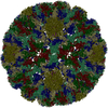
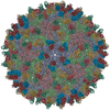
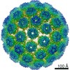
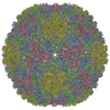
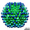
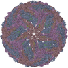
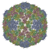
- PDB-5a1z: Cryo-EM structure of Dengue virus serotype 2 strain PVP94-07 comp... -
+ Open data

- Basic information
| Entry | Database: PDB / ID: 5a1z | ||||||
|---|---|---|---|---|---|---|---|
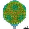
| Title | Cryo-EM structure of Dengue virus serotype 2 strain PVP94-07 complexed with human antibody 2D22 Fab at 37 degrees C | ||||||
 Components Components |
| ||||||
 Keywords Keywords |  VIRUS / DENGUE VIRUS / HUMAN ANTIBODY / CRYO-EM / NEUTRALIZATION VIRUS / DENGUE VIRUS / HUMAN ANTIBODY / CRYO-EM / NEUTRALIZATION | ||||||
| Function / homology |  Function and homology information Function and homology informationsymbiont-mediated suppression of host JAK-STAT cascade via inhibition of host TYK2 activity / symbiont-mediated suppression of host JAK-STAT cascade via inhibition of STAT2 activity / viral life cycle / ribonucleoside triphosphate phosphatase activity / viral capsid / double-stranded RNA binding / clathrin-dependent endocytosis of virus by host cell / mRNA (nucleoside-2'-O-)-methyltransferase activity / mRNA 5'-cap (guanine-N7-)-methyltransferase activity / RNA helicase activity ...symbiont-mediated suppression of host JAK-STAT cascade via inhibition of host TYK2 activity / symbiont-mediated suppression of host JAK-STAT cascade via inhibition of STAT2 activity / viral life cycle / ribonucleoside triphosphate phosphatase activity / viral capsid / double-stranded RNA binding / clathrin-dependent endocytosis of virus by host cell / mRNA (nucleoside-2'-O-)-methyltransferase activity / mRNA 5'-cap (guanine-N7-)-methyltransferase activity / RNA helicase activity / host cell endoplasmic reticulum membrane / membrane => GO:0016020 / protein dimerization activity / viral RNA genome replication / RNA-dependent RNA polymerase activity / serine-type endopeptidase activity / fusion of virus membrane with host endosome membrane / viral envelope / symbiont-mediated suppression of host type I interferon-mediated signaling pathway / host cell nucleus / structural molecule activity / virion attachment to host cell / virion membrane / proteolysis / extracellular region / ATP binding / membrane / metal ion bindingSimilarity search - Function | ||||||
| Biological species |  DENGUE VIRUS 2 DENGUE VIRUS 2 HOMO SAPIENS (human) HOMO SAPIENS (human) | ||||||
| Method | ELECTRON MICROSCOPY / single particle reconstruction / cryo EM / Resolution: 6.9 Å | ||||||
| Model type details | CA ATOMS ONLY, CHAIN A, C, E, B, D, F, G, I, K, H, J, L AUTHOR G.FIBRIANSAH,K.D.IBARRA,T.S.NG,S.A. ...CA ATOMS ONLY, CHAIN A, C, E, B, D, F, G, I, K, H, J, L AUTHOR G.FIBRIANSAH,K.D.IBARRA,T.S.NG,S.A.SMITH,J.L.TAN,X.N.LIM,J.S.G.OOI, | ||||||
 Authors Authors | Fibriansah, G. / Ibarra, K.D. / Ng, T.S. / Smith, S.A. / Tan, J.L. / Lim, X.N. / Ooi, J.S.G. / Kostyuchenko, V.A. / Wang, J. / de Silva, A.M. ...Fibriansah, G. / Ibarra, K.D. / Ng, T.S. / Smith, S.A. / Tan, J.L. / Lim, X.N. / Ooi, J.S.G. / Kostyuchenko, V.A. / Wang, J. / de Silva, A.M. / Harris, E. / Crowe Jr, J.E. / Lok, S.M. | ||||||
 Citation Citation | Journal: Science / Year: 2015 Title: DENGUE VIRUS. Cryo-EM structure of an antibody that neutralizes dengue virus type 2 by locking E protein dimers. Authors: Guntur Fibriansah / Kristie D Ibarra / Thiam-Seng Ng / Scott A Smith / Joanne L Tan / Xin-Ni Lim / Justin S G Ooi / Victor A Kostyuchenko / Jiaqi Wang / Aravinda M de Silva / Eva Harris / ...Authors: Guntur Fibriansah / Kristie D Ibarra / Thiam-Seng Ng / Scott A Smith / Joanne L Tan / Xin-Ni Lim / Justin S G Ooi / Victor A Kostyuchenko / Jiaqi Wang / Aravinda M de Silva / Eva Harris / James E Crowe / Shee-Mei Lok /   Abstract: There are four closely-related dengue virus (DENV) serotypes. Infection with one serotype generates antibodies that may cross-react and enhance infection with other serotypes in a secondary infection. ...There are four closely-related dengue virus (DENV) serotypes. Infection with one serotype generates antibodies that may cross-react and enhance infection with other serotypes in a secondary infection. We demonstrated that DENV serotype 2 (DENV2)-specific human monoclonal antibody (HMAb) 2D22 is therapeutic in a mouse model of antibody-enhanced severe dengue disease. We determined the cryo-electron microscopy (cryo-EM) structures of HMAb 2D22 complexed with two different DENV2 strains. HMAb 2D22 binds across viral envelope (E) proteins in the dimeric structure, which probably blocks the E protein reorganization required for virus fusion. HMAb 2D22 "locks" two-thirds of or all dimers on the virus surface, depending on the strain, but neutralizes these DENV2 strains with equal potency. The epitope defined by HMAb 2D22 is a potential target for vaccines and therapeutics. | ||||||
| History |
|
- Structure visualization
| Movie |
Movie viewer |
|---|---|
| Structure viewer | Molecule: MolmilJmol/JSmol |
- Downloads & links
-Download
| PDBx/mmCIF format | 5a1z.cif.gz | 78.5 KB | Display | PDBx/mmCIF format |
|---|---|---|---|---|
| PDB format | pdb5a1z.ent.gz | 51.1 KB | Display | PDB format |
| PDBx/mmJSON format | 5a1z.json.gz | Tree view | PDBx/mmJSON format | |
| Others |  Other downloads Other downloads |
-Validation report
| Arichive directory | https://data.pdbj.org/pub/pdb/validation_reports/a1/5a1zftp://data.pdbj.org/pub/pdb/validation_reports/a1/5a1z | HTTPS FTP |
|---|
-Related structure data
| Related structure data | 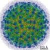 2996MC 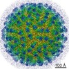 2967C  2968C 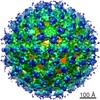 2969C  2997C 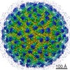 2998C 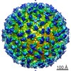 2999C  4uifC  4uihC M: map data used to model this data C: citing same article ( |
|---|---|
| Similar structure data |
-Links
 PDBj
PDBj


- Assembly
| Deposited unit | 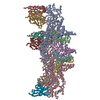
|
|---|---|
| 1 | x 60
|
| 2 |
|
| 3 | x 5
|
| 4 | x 6
|
| 5 | 
|
| Symmetry | Point symmetry: (Schoenflies symbol: I (icosahedral)) |
-Components
| #1: Protein | Viral envelope / E PROTEIN / Coordinate model: Cα atoms only Mass: 54258.551 Da / Num. of mol.: 3 Source method: isolated from a genetically manipulated source Source: (gene. exp.) DENGUE VIRUS 2 / Strain: PVP94-07Description: THE C6/36 CELLS WERE INFECTED WITH DENGUE VIRUS SEROTYPE 2 STRAIN PVP94/07 Cell line (production host): C6/36 / Production host:  AEDES ALBOPICTUS (Asian tiger mosquito) / References: UniProt: G9FRP5, UniProt: E0WXJ2*PLUS AEDES ALBOPICTUS (Asian tiger mosquito) / References: UniProt: G9FRP5, UniProt: E0WXJ2*PLUS#2: Protein | Glycoprotein / M PROTEIN / Coordinate model: Cα atoms onlyMass: 8109.491 Da / Num. of mol.: 3 / Fragment: RESIDUES 92-163 Source method: isolated from a genetically manipulated source Source: (gene. exp.) DENGUE VIRUS 2 / Strain: PVP94-07Description: THE C6/36 CELLS WERE INFECTED WITH DENGUE VIRUS SEROTYPE 2 STRAIN PVP94/07 Cell line (production host): C6/36 / Production host: AEDES ALBOPICTUS (Asian tiger mosquito) / References: UniProt: V5TI05, UniProt: E0WXJ2*PLUS#3: Antibody | Mass: 13805.458 Da / Num. of mol.: 3 / Source method: isolated from a natural source Details: 180 COPIES OF FAB 2D22 MOLECULE BIND TO VIRUS SURFACE Source: (natural) HOMO SAPIENS (human) / Cell: MEMORY B-CELLS#4: Antibody | Mass: 12090.306 Da / Num. of mol.: 3 / Source method: isolated from a natural source Details: 180 COPIES OF FAB 2D22 MOLECULE BIND TO VIRUS SURFACE Source: (natural) HOMO SAPIENS (human) / Cell: MEMORY B-CELLS |
|---|
-Experimental details
-Experiment
| Experiment | Method: ELECTRON MICROSCOPY |
|---|---|
| EM experiment | Aggregation state: PARTICLE / 3D reconstruction method: single particle reconstruction |
- Sample preparation
| Component | Name: DENGUE VIRUS SEROTYPE 2 STRAIN PVP94-07 (A CLINICAL ISOLATE) COMPLEXED WITH FAB FRAGMENTS OF HUMAN ANTIBODY 2D22. Type: COMPLEX Details: THE COMPLEX WAS INCUBATED AT 37 DEGREES C FOR 30 MIN |
|---|---|
| Buffer solution | Name: 10 MM TRIS-HCL PH 8.0, 120 MM NACL AND 1 MM EDTA / pH: 8 / Details: 10 MM TRIS-HCL PH 8.0, 120 MM NACL AND 1 MM EDTA |
| Specimen | Embedding applied: NO / Shadowing applied: NO / Staining applied: NO / Vitrification applied: YES |
| Specimen support | Details: HOLEY CARBON |
| Vitrification | Instrument: FEI VITROBOT MARK IV / Cryogen name: ETHANE Details: VITRIFICATION 1 -- CRYOGEN- ETHANE, HUMIDITY- 100, INSTRUMENT- FEI VITROBOT MARK IV, METHOD- BLOTTED WITH FILTER PAPER FOR 2 SECONDS PRIOR TO SNAP FREEZING, |
- Electron microscopy imaging
| Experimental equipment |  Model: Titan Krios / Image courtesy: FEI Company |
|---|---|
| Microscopy | Model: FEI TITAN KRIOS / Date: Feb 27, 2014 |
| Electron gun | Electron source: FIELD EMISSION GUN / Accelerating voltage: 300 kV / Illumination mode: SPOT SCAN |
| Electron lens | Mode: BRIGHT FIELDBright-field microscopy / Nominal magnification: 47000 X / Nominal defocus max: 4200 nm / Nominal defocus min: 1100 nm / Cs: 2.7 mm |
| Image recording | Electron dose: 18 e/Å2 / Film or detector model: FEI FALCON II (4k x 4k) |
| Image scans | Num. digital images: 177 |
- Processing
| EM software |
| ||||||||||||||||||||||||||||
|---|---|---|---|---|---|---|---|---|---|---|---|---|---|---|---|---|---|---|---|---|---|---|---|---|---|---|---|---|---|
| CTF correction | Details: EACH PARTICLE | ||||||||||||||||||||||||||||
| Symmetry | Point symmetry: I (icosahedral) | ||||||||||||||||||||||||||||
| 3D reconstruction | Method: CROSS-COMMON LINES / Resolution: 6.9 Å / Num. of particles: 3435 / Actual pixel size: 1.69 Å Details: SUBMISSION BASED ON EXPERIMENTAL DATA FROM EMDB EMD-2996. (DEPOSITION ID: 13351). Symmetry type: POINT | ||||||||||||||||||||||||||||
| Atomic model building | Protocol: FLEXIBLE FIT / Space: REAL / Target criteria: REAL-SPACE CORRELATION / Details: METHOD--FLEXIBLE REFINEMENT PROTOCOL--CRYO-EM | ||||||||||||||||||||||||||||
| Atomic model building | PDB-ID: 3J27 | ||||||||||||||||||||||||||||
| Refinement | Highest resolution: 6.9 Å | ||||||||||||||||||||||||||||
| Refinement step | Cycle: LAST / Highest resolution: 6.9 Å
|