 Movie
Movie Controller

[English] 日本語
 Yorodumi
- PDB-6i1o: Fab fragment of an antibody selective for wild-type alpha-1-antit... -
+ Open data

- Basic information
| Entry | Database: PDB / ID: 6i1o | |||||||||||||||
|---|---|---|---|---|---|---|---|---|---|---|---|---|---|---|---|---|
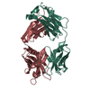
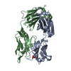
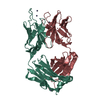
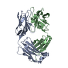
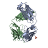
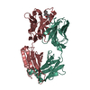
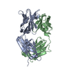
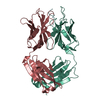
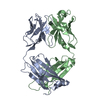
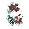
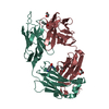
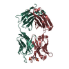
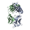
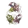
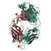
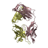
| Title | Fab fragment of an antibody selective for wild-type alpha-1-antitrypsin | |||||||||||||||
 Components Components |
| |||||||||||||||
 Keywords Keywords |  PROTEIN BINDING / Antibody fragment / Antitrypsin binding / Diagnostic / Monoclonal / selective / wild-type / Glu342 / E342 PROTEIN BINDING / Antibody fragment / Antitrypsin binding / Diagnostic / Monoclonal / selective / wild-type / Glu342 / E342 | |||||||||||||||
| Function / homology | Immunoglobulins / Immunoglobulin-like / Sandwich / Mainly Beta Function and homology information Function and homology information | |||||||||||||||
| Biological species |  Mus musculus (house mouse) Mus musculus (house mouse) | |||||||||||||||
| Method | X-RAY DIFFRACTION / SYNCHROTRON / MOLECULAR REPLACEMENT / Resolution: 1.93 Å | |||||||||||||||
 Authors Authors | Laffranchi, M. / Elliston, E.L.K. / Miranda, E. / Perez, J. / Fra, A. / Lomas, D.A. / Irving, J.A. | |||||||||||||||
| Funding support |  United Kingdom, United Kingdom,  Italy, Italy,  United States, 4items United States, 4items
| |||||||||||||||
 Citation Citation | Journal: JCI Insight / Year: 2020 Title: Intrahepatic heteropolymerization of M and Z alpha-1-antitrypsin. Authors: Laffranchi, M. / Elliston, E.L. / Miranda, E. / Perez, J. / Ronzoni, R. / Jagger, A.M. / Heyer-Chauhan, N. / Brantly, M.L. / Fra, A. / Lomas, D.A. / Irving, J.A. | |||||||||||||||
| History |
|
- Structure visualization
| Structure viewer | Molecule: MolmilJmol/JSmol |
|---|
- Downloads & links
-Download
| PDBx/mmCIF format | 6i1o.cif.gz | 115.7 KB | Display | PDBx/mmCIF format |
|---|---|---|---|---|
| PDB format | pdb6i1o.ent.gz | 77.8 KB | Display | PDB format |
| PDBx/mmJSON format | 6i1o.json.gz | Tree view | PDBx/mmJSON format | |
| Others |  Other downloads Other downloads |
-Validation report
| Arichive directory | https://data.pdbj.org/pub/pdb/validation_reports/i1/6i1oftp://data.pdbj.org/pub/pdb/validation_reports/i1/6i1o | HTTPS FTP |
|---|
-Related structure data
| Related structure data | 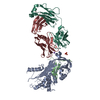 6i3zC 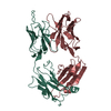 1ae6S S: Starting model for refinement C: citing same article ( |
|---|---|
| Similar structure data |


-Links
 PDBj
PDBj

- Assembly
| Deposited unit | 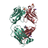
| ||||||||||||
|---|---|---|---|---|---|---|---|---|---|---|---|---|---|
| 1 |
| ||||||||||||
| Unit cell |
| ||||||||||||
| Components on special symmetry positions |
|
-Components
-Antibody , 2 types, 2 molecules HL
| #1: Antibody | Mass: 23129.936 Da / Num. of mol.: 1 / Source method: isolated from a natural source / Source: (natural) Mus musculus (house mouse) / Cell line: Hybridoma / Organ: Spleen / Variant: BALB/c |
|---|---|
| #2: Antibody | Mass: 23811.256 Da / Num. of mol.: 1 / Source method: isolated from a natural source / Source: (natural) Mus musculus (house mouse) / Cell line: Hybridoma / Organ: Spleen / Variant: BALB/c |
-Non-polymers , 4 types, 372 molecules 


| #3: Chemical | Sulfate Mass: 96.063 Da / Num. of mol.: 2 / Source method: obtained synthetically / Formula: SO4 Mass: 96.063 Da / Num. of mol.: 2 / Source method: obtained synthetically / Formula: SO4#4: Chemical | ChemComp-NA /  Mass: 22.990 Da / Num. of mol.: 4 / Source method: obtained synthetically / Formula: Na Mass: 22.990 Da / Num. of mol.: 4 / Source method: obtained synthetically / Formula: Na#5: Chemical | ChemComp-BTB / | Bis-tris methane Mass: 209.240 Da / Num. of mol.: 1 / Source method: obtained synthetically / Formula: C8H19NO5 / Comment: pH buffer*YM Mass: 209.240 Da / Num. of mol.: 1 / Source method: obtained synthetically / Formula: C8H19NO5 / Comment: pH buffer*YM#6: Water | ChemComp-HOH / | WaterMass: 18.015 Da / Num. of mol.: 365 / Source method: isolated from a natural source / Formula: H2O |
|---|
-Experimental details
-Experiment
| Experiment | Method: X-RAY DIFFRACTION / Number of used crystals: 1 |
|---|
- Sample preparation
| Crystal | Density Matthews: 2.44 Å3/Da / Density % sol: 49.68 % / Description: Bladed |
|---|---|
| Crystal grow | Temperature: 293 K / Method: vapor diffusion, hanging drop / pH: 6 Details: 20% PEG 3350, 0.2M Ammonium Sulfate, 0.1M Bis-Tris pH 6.0 |
-Data collection
| Diffraction | Mean temperature: 100 K / Ambient temp details: Cryostream / Serial crystal experiment: N | ||||||||||||||||||||||||||||||||||||||||||||||||||||||||||||||||||||||||||||||||||||||||||||||||||||||||||||||||||||||||||||||||||||||||||||||||||||||||||||||||||||||||||||||||||||||||||||||||||||||||||||||||||
|---|---|---|---|---|---|---|---|---|---|---|---|---|---|---|---|---|---|---|---|---|---|---|---|---|---|---|---|---|---|---|---|---|---|---|---|---|---|---|---|---|---|---|---|---|---|---|---|---|---|---|---|---|---|---|---|---|---|---|---|---|---|---|---|---|---|---|---|---|---|---|---|---|---|---|---|---|---|---|---|---|---|---|---|---|---|---|---|---|---|---|---|---|---|---|---|---|---|---|---|---|---|---|---|---|---|---|---|---|---|---|---|---|---|---|---|---|---|---|---|---|---|---|---|---|---|---|---|---|---|---|---|---|---|---|---|---|---|---|---|---|---|---|---|---|---|---|---|---|---|---|---|---|---|---|---|---|---|---|---|---|---|---|---|---|---|---|---|---|---|---|---|---|---|---|---|---|---|---|---|---|---|---|---|---|---|---|---|---|---|---|---|---|---|---|---|---|---|---|---|---|---|---|---|---|---|---|---|---|---|---|---|
| Diffraction source | Source: SYNCHROTRON / Site: Diamond / Beamline: I03 / Wavelength: 0.9793 Å | ||||||||||||||||||||||||||||||||||||||||||||||||||||||||||||||||||||||||||||||||||||||||||||||||||||||||||||||||||||||||||||||||||||||||||||||||||||||||||||||||||||||||||||||||||||||||||||||||||||||||||||||||||
| Detector | Type: DECTRIS PILATUS3 6M / Detector: PIXEL / Date: Jul 8, 2018 | ||||||||||||||||||||||||||||||||||||||||||||||||||||||||||||||||||||||||||||||||||||||||||||||||||||||||||||||||||||||||||||||||||||||||||||||||||||||||||||||||||||||||||||||||||||||||||||||||||||||||||||||||||
| Radiation | Monochromator: Double crystal / Protocol: SINGLE WAVELENGTH / Monochromatic (M) / Laue (L): M / Scattering type: x-ray | ||||||||||||||||||||||||||||||||||||||||||||||||||||||||||||||||||||||||||||||||||||||||||||||||||||||||||||||||||||||||||||||||||||||||||||||||||||||||||||||||||||||||||||||||||||||||||||||||||||||||||||||||||
| Radiation wavelength | Wavelength: 0.9793 Å / Relative weight: 1 | ||||||||||||||||||||||||||||||||||||||||||||||||||||||||||||||||||||||||||||||||||||||||||||||||||||||||||||||||||||||||||||||||||||||||||||||||||||||||||||||||||||||||||||||||||||||||||||||||||||||||||||||||||
| Reflection | Resolution: 1.93→85.91 Å / Num. obs: 665286 / % possible obs: 100 % / Redundancy: 19.2 % / Biso Wilson estimate: 29.38 Å2 / CC1/2: 0.999 / Rmerge(I) obs: 0.186 / Rpim(I) all: 0.043 / Rrim(I) all: 0.191 / Χ2: 0.95 / Net I/σ(I): 11.6 | ||||||||||||||||||||||||||||||||||||||||||||||||||||||||||||||||||||||||||||||||||||||||||||||||||||||||||||||||||||||||||||||||||||||||||||||||||||||||||||||||||||||||||||||||||||||||||||||||||||||||||||||||||
| Reflection shell | Diffraction-ID: 1
|
- Processing
| Software |
| |||||||||||||||||||||||||||||||||||||||||||||||||||||||||||||||||||||||||||||||||||||||||||
|---|---|---|---|---|---|---|---|---|---|---|---|---|---|---|---|---|---|---|---|---|---|---|---|---|---|---|---|---|---|---|---|---|---|---|---|---|---|---|---|---|---|---|---|---|---|---|---|---|---|---|---|---|---|---|---|---|---|---|---|---|---|---|---|---|---|---|---|---|---|---|---|---|---|---|---|---|---|---|---|---|---|---|---|---|---|---|---|---|---|---|---|---|
| Refinement | Method to determine structure: MOLECULAR REPLACEMENT Starting model: 1AE6 Resolution: 1.93→85.91 Å / SU ML: 0.2428 / Cross valid method: FREE R-VALUE / σ(F): 1.34 / Phase error: 24.3933
| |||||||||||||||||||||||||||||||||||||||||||||||||||||||||||||||||||||||||||||||||||||||||||
| Solvent computation | Shrinkage radii: 0.9 Å / VDW probe radii: 1.11 Å | |||||||||||||||||||||||||||||||||||||||||||||||||||||||||||||||||||||||||||||||||||||||||||
| Displacement parameters | Biso mean: 33.38 Å2 | |||||||||||||||||||||||||||||||||||||||||||||||||||||||||||||||||||||||||||||||||||||||||||
| Refinement step | Cycle: LAST / Resolution: 1.93→85.91 Å
| |||||||||||||||||||||||||||||||||||||||||||||||||||||||||||||||||||||||||||||||||||||||||||
| Refine LS restraints |
| |||||||||||||||||||||||||||||||||||||||||||||||||||||||||||||||||||||||||||||||||||||||||||
| LS refinement shell |
|
