 Movie
Movie Controller


+ Open data

- Basic information
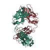
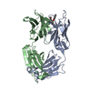
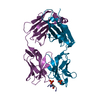
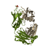
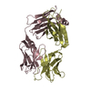
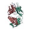
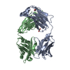
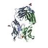
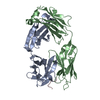
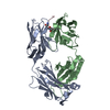
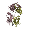
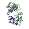
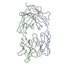
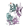
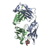
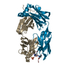
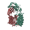
| Entry | Database: PDB / ID: 4x7t | ||||||||||||
|---|---|---|---|---|---|---|---|---|---|---|---|---|---|
| Title | Structure of Omalizumab Fab fragment, crystal form 2 | ||||||||||||
 Components Components |
| ||||||||||||
 Keywords Keywords |  IMMUNE SYSTEM / Antibody Fab Fragment / Anti-IgE antibody / Anti-inflammatory IMMUNE SYSTEM / Antibody Fab Fragment / Anti-IgE antibody / Anti-inflammatory | ||||||||||||
| Function / homology | Immunoglobulins / Immunoglobulin-like / Sandwich / Mainly Beta Function and homology information Function and homology information | ||||||||||||
| Biological species |  Homo sapiens (human) Homo sapiens (human) | ||||||||||||
| Method | X-RAY DIFFRACTION / SYNCHROTRON / MOLECULAR REPLACEMENT / Resolution: 3 Å | ||||||||||||
 Authors Authors | Jensen, R.K. / Andersen, G.R. | ||||||||||||
| Funding support |  Denmark, 3items Denmark, 3items
| ||||||||||||
 Citation Citation | Journal: Acta Crystallogr.,Sect.F / Year: 2015 Title: Structure of the omalizumab Fab Authors: Jensen, R.K. / Plum, M. / Tjerrild, L. / Jakob, T. / Spillner, E. / Andersen, G.R. | ||||||||||||
| History |
|
- Structure visualization
| Structure viewer | Molecule: MolmilJmol/JSmol |
|---|
- Downloads & links
-Download
| PDBx/mmCIF format | 4x7t.cif.gz | 344.6 KB | Display | PDBx/mmCIF format |
|---|---|---|---|---|
| PDB format | pdb4x7t.ent.gz | 282.9 KB | Display | PDB format |
| PDBx/mmJSON format | 4x7t.json.gz | Tree view | PDBx/mmJSON format | |
| Others |  Other downloads Other downloads |
-Validation report
| Arichive directory | https://data.pdbj.org/pub/pdb/validation_reports/x7/4x7tftp://data.pdbj.org/pub/pdb/validation_reports/x7/4x7t | HTTPS FTP |
|---|
-Related structure data
| Related structure data |  4x7sSC S: Starting model for refinement C: citing same article ( |
|---|---|
| Similar structure data |


-Links
 PDBj
PDBj

- Assembly
| Deposited unit | 
| ||||||||
|---|---|---|---|---|---|---|---|---|---|
| 1 | 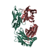
| ||||||||
| 2 | 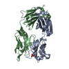
| ||||||||
| Unit cell |
|
-Components
| #1: Antibody | Mass: 23653.387 Da / Num. of mol.: 2 Source method: isolated from a genetically manipulated source Source: (gene. exp.) Homo sapiens (human) / Production host:   Cricetulus griseus (Chinese hamster) Cricetulus griseus (Chinese hamster)#2: Antibody | Mass: 23922.287 Da / Num. of mol.: 2 Source method: isolated from a genetically manipulated source Source: (gene. exp.) Homo sapiens (human) / Production host: Cricetulus griseus (Chinese hamster)#3: Chemical | Sulfate  Mass: 96.063 Da / Num. of mol.: 2 / Source method: obtained synthetically / Formula: SO4 Mass: 96.063 Da / Num. of mol.: 2 / Source method: obtained synthetically / Formula: SO4 |
|---|
-Experimental details
-Experiment
| Experiment | Method: X-RAY DIFFRACTION |
|---|
- Sample preparation
| Crystal | Density Matthews: 2.57 Å3/Da / Density % sol: 52.18 % |
|---|---|
| Crystal grow | Temperature: 293 K / Method: vapor diffusion, sitting drop / Details: HEPES, Polyvinyl Pyrrolidone K15, Lithium Sulphate |
-Data collection
| Diffraction | Mean temperature: 100 K |
|---|---|
| Diffraction source | Source: SYNCHROTRON / Site: PETRA III, EMBL c/o DESY  / Beamline: P14 (MX2) / Wavelength: 0.97551 Å / Beamline: P14 (MX2) / Wavelength: 0.97551 Å |
| Detector | Type: DECTRIS PILATUS 6M-F / Detector: PIXEL / Date: Nov 29, 2013 |
| Radiation | Protocol: SINGLE WAVELENGTH / Monochromatic (M) / Laue (L): M / Scattering type: x-ray |
| Radiation wavelength | Wavelength: 0.97551 Å / Relative weight: 1 |
| Reflection | Resolution: 3→45.41 Å / Num. obs: 19417 / % possible obs: 99.37 % / Redundancy: 3.4 % / Rsym value: 0.202 / Net I/σ(I): 5.8 |
| Reflection shell | Resolution: 3→3.107 Å / Redundancy: 3.2 % / Mean I/σ(I) obs: 1.11 / % possible all: 99.32 |
- Processing
| Software |
| |||||||||||||||||||||||||||||||||||||||||||||||||||||||||||||||||||||||||||||||||||||||||||||||||||||||||
|---|---|---|---|---|---|---|---|---|---|---|---|---|---|---|---|---|---|---|---|---|---|---|---|---|---|---|---|---|---|---|---|---|---|---|---|---|---|---|---|---|---|---|---|---|---|---|---|---|---|---|---|---|---|---|---|---|---|---|---|---|---|---|---|---|---|---|---|---|---|---|---|---|---|---|---|---|---|---|---|---|---|---|---|---|---|---|---|---|---|---|---|---|---|---|---|---|---|---|---|---|---|---|---|---|---|---|
| Refinement | Method to determine structure: MOLECULAR REPLACEMENT Starting model: 4X7S Resolution: 3→45.408 Å / SU ML: 0.41 / Cross valid method: THROUGHOUT / σ(F): 1.37 / Phase error: 25.32 / Stereochemistry target values: ML
| |||||||||||||||||||||||||||||||||||||||||||||||||||||||||||||||||||||||||||||||||||||||||||||||||||||||||
| Solvent computation | Shrinkage radii: 0.9 Å / VDW probe radii: 1.11 Å / Solvent model: FLAT BULK SOLVENT MODEL | |||||||||||||||||||||||||||||||||||||||||||||||||||||||||||||||||||||||||||||||||||||||||||||||||||||||||
| Refinement step | Cycle: LAST / Resolution: 3→45.408 Å
| |||||||||||||||||||||||||||||||||||||||||||||||||||||||||||||||||||||||||||||||||||||||||||||||||||||||||
| Refine LS restraints |
| |||||||||||||||||||||||||||||||||||||||||||||||||||||||||||||||||||||||||||||||||||||||||||||||||||||||||
| LS refinement shell |
| |||||||||||||||||||||||||||||||||||||||||||||||||||||||||||||||||||||||||||||||||||||||||||||||||||||||||
| Refinement TLS params. | Method: refined / Origin x: 14.6159 Å / Origin y: -3.0265 Å / Origin z: 18.1444 Å
| |||||||||||||||||||||||||||||||||||||||||||||||||||||||||||||||||||||||||||||||||||||||||||||||||||||||||
| Refinement TLS group | Selection details: all |
