 Movie
Movie Controller

[English] 日本語
 Yorodumi
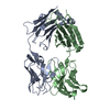
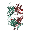
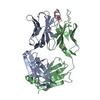
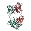
- PDB-5xhf: Crystal structure of Trastuzumab Fab fragment bearing p-azido-L-p... -
+ Open data

- Basic information
| Entry | Database: PDB / ID: 5xhf | ||||||
|---|---|---|---|---|---|---|---|
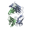
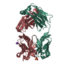
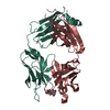
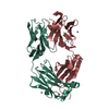
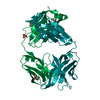
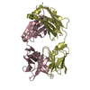
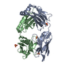
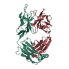
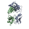
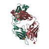
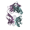
| Title | Crystal structure of Trastuzumab Fab fragment bearing p-azido-L-phenylalanine | ||||||
 Components Components |
| ||||||
 Keywords Keywords |  IMMUNE SYSTEM / FAB IMMUNE SYSTEM / FAB | ||||||
| Function / homology | Immunoglobulins / Immunoglobulin-like / Sandwich / Mainly Beta Function and homology information Function and homology information | ||||||
| Biological species |  MUS MUSCULUS (house mouse) MUS MUSCULUS (house mouse) | ||||||
| Method | X-RAY DIFFRACTION / SYNCHROTRON / MOLECULAR REPLACEMENT / Resolution: 3.205 Å | ||||||
 Authors Authors | Kuratani, M. / Yanagisawa, T. / Sakamoto, K. / Yokoyama, S. | ||||||
 Citation Citation | Journal: Bioconjug. Chem. / Year: 2017 Title: Extensive Survey of Antibody Invariant Positions for Efficient Chemical Conjugation Using Expanded Genetic Codes. Authors: Kato, A. / Kuratani, M. / Yanagisawa, T. / Ohtake, K. / Hayashi, A. / Amano, Y. / Kimura, K. / Yokoyama, S. / Sakamoto, K. / Shiraishi, Y. | ||||||
| History |
|
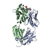
- Structure visualization
| Structure viewer | Molecule: MolmilJmol/JSmol |
|---|
- Downloads & links
-Download
| PDBx/mmCIF format | 5xhf.cif.gz | 172.9 KB | Display | PDBx/mmCIF format |
|---|---|---|---|---|
| PDB format | pdb5xhf.ent.gz | 137.3 KB | Display | PDB format |
| PDBx/mmJSON format | 5xhf.json.gz | Tree view | PDBx/mmJSON format | |
| Others |  Other downloads Other downloads |
-Validation report
| Arichive directory | https://data.pdbj.org/pub/pdb/validation_reports/xh/5xhfftp://data.pdbj.org/pub/pdb/validation_reports/xh/5xhf | HTTPS FTP |
|---|
-Related structure data
| Related structure data |  5xhgC 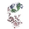 1n8zS C: citing same article ( S: Starting model for refinement |
|---|---|
| Similar structure data |

-Links
 PDBj
PDBj

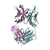
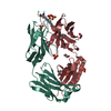
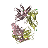
- Assembly
| Deposited unit | 
| ||||||||
|---|---|---|---|---|---|---|---|---|---|
| 1 | 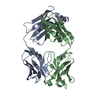
| ||||||||
| 2 | 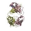
| ||||||||
| Unit cell |
|
-Components
| #1: Antibody | Mass: 23527.096 Da / Num. of mol.: 2 Source method: isolated from a genetically manipulated source Source: (gene. exp.) MUS MUSCULUS (house mouse) / Description: humanized mouseProduction host:  Escherichia coli str. K-12 substr. W3110 (bacteria) Escherichia coli str. K-12 substr. W3110 (bacteria)#2: Antibody | Mass: 23425.180 Da / Num. of mol.: 2 Source method: isolated from a genetically manipulated source Source: (gene. exp.) MUS MUSCULUS (house mouse) / Description: humanized mouseProduction host: Escherichia coli str. K-12 substr. W3110 (bacteria) |
|---|
-Experimental details
-Experiment
| Experiment | Method: X-RAY DIFFRACTION / Number of used crystals: 1 |
|---|
- Sample preparation
| Crystal | Density Matthews: 2.51 Å3/Da / Density % sol: 51.01 % / Description: THE FILE CONTAINS FRIEDEL PAIRS. |
|---|---|
| Crystal grow | Temperature: 293 K / Method: vapor diffusion, sitting drop / pH: 6.5 / Details: PEG 20000, MES-Na |
-Data collection
| Diffraction | Mean temperature: 99 K |
|---|---|
| Diffraction source | Source: SYNCHROTRON / Site: NSRRC  / Beamline: TPS 05A / Wavelength: 1 Å / Beamline: TPS 05A / Wavelength: 1 Å |
| Detector | Type: RAYONIX MX300-HS / Detector: CCD / Date: Mar 13, 2017 |
| Radiation | Protocol: SINGLE WAVELENGTH / Monochromatic (M) / Laue (L): M / Scattering type: x-ray |
| Radiation wavelength | Wavelength: 1 Å / Relative weight: 1 |
| Reflection | Resolution: 3.2→50 Å / Num. obs: 14758 / % possible obs: 95.7 % / Redundancy: 1.3 % / Net I/σ(I): 2.45 |
- Processing
| Software |
| ||||||||||||||||||||||||||||||||||||||||||||||||||||||||||||||||||||||||||||||||||||
|---|---|---|---|---|---|---|---|---|---|---|---|---|---|---|---|---|---|---|---|---|---|---|---|---|---|---|---|---|---|---|---|---|---|---|---|---|---|---|---|---|---|---|---|---|---|---|---|---|---|---|---|---|---|---|---|---|---|---|---|---|---|---|---|---|---|---|---|---|---|---|---|---|---|---|---|---|---|---|---|---|---|---|---|---|---|
| Refinement | Method to determine structure: MOLECULAR REPLACEMENT Starting model: 1N8Z Resolution: 3.205→42.443 Å / SU ML: 0.47 / Cross valid method: FREE R-VALUE / σ(F): 1.98 / Phase error: 32.76 / Details: THE FILE CONTAINS FRIEDEL PAIRS.
| ||||||||||||||||||||||||||||||||||||||||||||||||||||||||||||||||||||||||||||||||||||
| Solvent computation | Shrinkage radii: 0.9 Å / VDW probe radii: 1.11 Å | ||||||||||||||||||||||||||||||||||||||||||||||||||||||||||||||||||||||||||||||||||||
| Refinement step | Cycle: LAST / Resolution: 3.205→42.443 Å
| ||||||||||||||||||||||||||||||||||||||||||||||||||||||||||||||||||||||||||||||||||||
| Refine LS restraints |
| ||||||||||||||||||||||||||||||||||||||||||||||||||||||||||||||||||||||||||||||||||||
| LS refinement shell |
|
