 Movie
Movie Controller

[English] 日本語
 Yorodumi
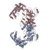
- PDB-1n8z: Crystal structure of extracellular domain of human HER2 complexed... -
+ Open data

- Basic information
| Entry | Database: PDB / ID: 1n8z | ||||||
|---|---|---|---|---|---|---|---|
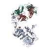
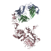
| Title | Crystal structure of extracellular domain of human HER2 complexed with Herceptin Fab | ||||||
 Components Components |
| ||||||
 Keywords Keywords |  TRANSFERASE / tyrosin kinase receptor / cell surface receptor TRANSFERASE / tyrosin kinase receptor / cell surface receptor | ||||||
| Function / homology |  Function and homology information Function and homology informationnegative regulation of immature T cell proliferation in thymus / ERBB3:ERBB2 complex / ERBB2-ERBB4 signaling pathway / GRB7 events in ERBB2 signaling / immature T cell proliferation in thymus / RNA polymerase I core binding / regulation of microtubule-based process / ErbB-3 class receptor binding / semaphorin receptor complex / Sema4D induced cell migration and growth-cone collapse ...negative regulation of immature T cell proliferation in thymus / ERBB3:ERBB2 complex / ERBB2-ERBB4 signaling pathway / GRB7 events in ERBB2 signaling / immature T cell proliferation in thymus / RNA polymerase I core binding / regulation of microtubule-based process / ErbB-3 class receptor binding / semaphorin receptor complex / Sema4D induced cell migration and growth-cone collapse / motor neuron axon guidance / neurotransmitter receptor localization to postsynaptic specialization membrane / PLCG1 events in ERBB2 signaling / ERBB2-EGFR signaling pathway / neuromuscular junction development / positive regulation of Rho protein signal transduction / ERBB2 Activates PTK6 Signaling / Drug-mediated inhibition of ERBB2 signaling / Resistance of ERBB2 KD mutants to trastuzumab / Resistance of ERBB2 KD mutants to sapitinib / Resistance of ERBB2 KD mutants to tesevatinib / Resistance of ERBB2 KD mutants to neratinib / Resistance of ERBB2 KD mutants to osimertinib / Resistance of ERBB2 KD mutants to afatinib / Resistance of ERBB2 KD mutants to AEE788 / Resistance of ERBB2 KD mutants to lapatinib / Drug resistance in ERBB2 TMD/JMD mutants / enzyme-linked receptor protein signaling pathway / positive regulation of transcription by RNA polymerase I / ERBB2-ERBB3 signaling pathway / oligodendrocyte differentiation / ERBB2 Regulates Cell Motility / semaphorin-plexin signaling pathway / PI3K events in ERBB2 signaling / positive regulation of cell adhesion / positive regulation of protein targeting to membrane / regulation of angiogenesis / coreceptor activity / Schwann cell development / Signaling by ERBB2 / cellular response to epidermal growth factor stimulus / myelination / Downregulation of ERBB2:ERBB3 signaling / GRB2 events in ERBB2 signaling / TFAP2 (AP-2) family regulates transcription of growth factors and their receptors / transmembrane receptor protein tyrosine kinase activity / SHC1 events in ERBB2 signaling / Constitutive Signaling by Overexpressed ERBB2 / neurogenesis / basal plasma membrane / regulation of ERK1 and ERK2 cascade / phosphatidylinositol 3-kinase/protein kinase B signal transduction / positive regulation of translation / positive regulation of epithelial cell proliferation / cell surface receptor protein tyrosine kinase signaling pathway / Signaling by ERBB2 TMD/JMD mutants / positive regulation of MAP kinase activity / wound healing / neuromuscular junction / Signaling by ERBB2 ECD mutants / neuron differentiation / Signaling by ERBB2 KD Mutants / receptor protein-tyrosine kinase / receptor tyrosine kinase binding / cellular response to growth factor stimulus / Downregulation of ERBB2 signaling / ruffle membrane / peptidyl-tyrosine phosphorylation / Constitutive Signaling by Aberrant PI3K in Cancer / transmembrane signaling receptor activity / PIP3 activates AKT signaling / myelin sheath / presynaptic membrane / heart development / PI5P, PP2A and IER3 Regulate PI3K/AKT Signaling / RAF/MAP kinase cascade / positive regulation of cell growth / basolateral plasma membrane / protein tyrosine kinase activity / positive regulation of MAPK cascade / early endosome / cell surface receptor signaling pathway / receptor complex / endosome membrane / intracellular signal transduction / apical plasma membrane / positive regulation of protein phosphorylation / protein heterodimerization activity / protein phosphorylation / signaling receptor binding / positive regulation of cell population proliferation / negative regulation of apoptotic process / perinuclear region of cytoplasm / signal transduction / nucleoplasm / ATP binding / membrane / identical protein binding / nucleus / plasma membraneSimilarity search - Function | ||||||
| Biological species |  Mus musculus (house mouse) Mus musculus (house mouse) Homo sapiens (human) Homo sapiens (human) | ||||||
| Method | X-RAY DIFFRACTION / SYNCHROTRON / MOLECULAR REPLACEMENT / Resolution: 2.52 Å | ||||||
 Authors Authors | Cho, H.-S. / Mason, K. / Ramyar, K.X. / Stanley, A.M. / Gabelli, S.B. / Denney Jr., D.W. / Leahy, D.J. | ||||||
 Citation Citation | Journal: Nature / Year: 2003 Title: Structure of the Extracellular Region of HER2 Alone and in Complex with the Herceptin Fab Authors: Cho, H.-S. / Mason, K. / Ramyar, K.X. / Stanley, A.M. / Gabelli, S.B. / Denney Jr., D.W. / Leahy, D.J. | ||||||
| History |
| ||||||
| Remark 999 | SEQUENCE The sequences of Herseptin Fab light chain (molecule A) and heavy chain (molecule B) were ...SEQUENCE The sequences of Herseptin Fab light chain (molecule A) and heavy chain (molecule B) were not deposited into any database. |
- Structure visualization
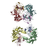
| Structure viewer | Molecule: MolmilJmol/JSmol |
|---|
- Downloads & links
-Download
| PDBx/mmCIF format | 1n8z.cif.gz | 206.8 KB | Display | PDBx/mmCIF format |
|---|---|---|---|---|
| PDB format | pdb1n8z.ent.gz | 162.9 KB | Display | PDB format |
| PDBx/mmJSON format | 1n8z.json.gz | Tree view | PDBx/mmJSON format | |
| Others |  Other downloads Other downloads |
-Validation report
| Arichive directory | https://data.pdbj.org/pub/pdb/validation_reports/n8/1n8zftp://data.pdbj.org/pub/pdb/validation_reports/n8/1n8z | HTTPS FTP |
|---|
-Related structure data
| Related structure data | 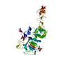 1n8yC 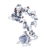 1m6bS C: citing same article ( S: Starting model for refinement |
|---|---|
| Similar structure data |


-Links
 PDBj
PDBj


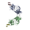
- Assembly
| Deposited unit | 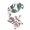
| ||||||||
|---|---|---|---|---|---|---|---|---|---|
| 1 |
| ||||||||
| Unit cell |
|
-Components
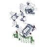
-Antibody , 2 types, 2 molecules AB
| #1: Antibody | Mass: 23466.031 Da / Num. of mol.: 1 Source method: isolated from a genetically manipulated source Source: (gene. exp.) Mus musculus, Homo sapiens |
|---|---|
| #2: Antibody | Mass: 23425.180 Da / Num. of mol.: 1 Source method: isolated from a genetically manipulated source Source: (gene. exp.) Mus musculus, Homo sapiens |
-Protein / Sugars , 2 types, 3 molecules C
| #3: Protein | / Tyrosine kinase-type cell surface receptor HER2 Mass: 66989.109 Da / Num. of mol.: 1 / Fragment: extracellular domain Source method: isolated from a genetically manipulated source Source: (gene. exp.) Homo sapiens (human) / Gene: HER2(erbB2) / Plasmid: pSGHV0 / Production host:  Cricetulus griseus (Chinese hamster) / Strain (production host): CHO(Lec1) / References: UniProt: P04626, EC: 2.7.1.112 Cricetulus griseus (Chinese hamster) / Strain (production host): CHO(Lec1) / References: UniProt: P04626, EC: 2.7.1.112 |
|---|---|
| #4: Sugar | N-Acetylglucosamine Type: D-saccharide, beta linking / Mass: 221.208 Da / Num. of mol.: 2 Type: D-saccharide, beta linking / Mass: 221.208 Da / Num. of mol.: 2Source method: isolated from a genetically manipulated source Formula: C8H15NO6 |
-Non-polymers , 2 types, 80 molecules 

| #5: Chemical | ChemComp-SO4 / Sulfate Mass: 96.063 Da / Num. of mol.: 1 / Source method: obtained synthetically / Formula: SO4 Mass: 96.063 Da / Num. of mol.: 1 / Source method: obtained synthetically / Formula: SO4 |
|---|---|
| #6: Water | ChemComp-HOH / WaterMass: 18.015 Da / Num. of mol.: 79 / Source method: isolated from a natural source / Formula: H2O |
-Experimental details
-Experiment
| Experiment | Method: X-RAY DIFFRACTION / Number of used crystals: 1 |
|---|
- Sample preparation
| Crystal | Density Matthews: 2.89 Å3/Da / Density % sol: 57.13 % | ||||||||||||||||||||||||||||||||||||
|---|---|---|---|---|---|---|---|---|---|---|---|---|---|---|---|---|---|---|---|---|---|---|---|---|---|---|---|---|---|---|---|---|---|---|---|---|---|
| Crystal grow | Temperature: 298 K / Method: vapor diffusion, hanging drop / pH: 6.5 Details: 20-30% PEG 5000 MME, 0.1M MES pH6.5, 0.2M ammonium sulfate, VAPOR DIFFUSION, HANGING DROP, temperature 298K | ||||||||||||||||||||||||||||||||||||
| Crystal grow | *PLUS pH: 7.5 | ||||||||||||||||||||||||||||||||||||
| Components of the solutions | *PLUS
|
-Data collection
| Diffraction | Mean temperature: 110 K |
|---|---|
| Diffraction source | Source: SYNCHROTRON / Site: NSLS  / Beamline: X4A / Beamline: X4A |
| Detector | Type: ADSC QUANTUM 4 / Detector: CCD |
| Radiation | Protocol: SINGLE WAVELENGTH / Monochromatic (M) / Laue (L): M / Scattering type: x-ray |
| Radiation wavelength | Relative weight: 1 |
| Reflection | Resolution: 2.5→20 Å / Num. all: 44400 / Num. obs: 42223 / % possible obs: 96.4 % / Observed criterion σ(F): 1 / Observed criterion σ(I): 1 / Redundancy: 3.9 % |
| Reflection shell | Resolution: 2.52→2.58 Å / % possible all: 90 |
| Reflection | *PLUS Lowest resolution: 30 Å / Num. measured all: 164522 / Rmerge(I) obs: 0.068 |
| Reflection shell | *PLUS % possible obs: 82.4 % / Rmerge(I) obs: 0.51 / Mean I/σ(I) obs: 1.9 |
- Processing
| Software |
| ||||||||||||||||||||||||||||||||||||||||||||||||||||||||||||||||||||||||||||||||||||||||||||||||||||||||||||||||||||||||||||||||||
|---|---|---|---|---|---|---|---|---|---|---|---|---|---|---|---|---|---|---|---|---|---|---|---|---|---|---|---|---|---|---|---|---|---|---|---|---|---|---|---|---|---|---|---|---|---|---|---|---|---|---|---|---|---|---|---|---|---|---|---|---|---|---|---|---|---|---|---|---|---|---|---|---|---|---|---|---|---|---|---|---|---|---|---|---|---|---|---|---|---|---|---|---|---|---|---|---|---|---|---|---|---|---|---|---|---|---|---|---|---|---|---|---|---|---|---|---|---|---|---|---|---|---|---|---|---|---|---|---|---|---|---|
| Refinement | Method to determine structure: MOLECULAR REPLACEMENT Starting model: PDB 1M6B Resolution: 2.52→20 Å / Cor.coef. Fo:Fc: 0.926 / Cor.coef. Fo:Fc free: 0.871 / SU B: 13.011 / SU ML: 0.279 / Cross valid method: THROUGHOUT / σ(F): 1 / σ(I): 1 / ESU R: 0.671 / ESU R Free: 0.342
| ||||||||||||||||||||||||||||||||||||||||||||||||||||||||||||||||||||||||||||||||||||||||||||||||||||||||||||||||||||||||||||||||||
| Solvent computation | Ion probe radii: 0.8 Å / Shrinkage radii: 0.8 Å / VDW probe radii: 1.4 Å / Solvent model: BABINET MODEL WITH MASK | ||||||||||||||||||||||||||||||||||||||||||||||||||||||||||||||||||||||||||||||||||||||||||||||||||||||||||||||||||||||||||||||||||
| Displacement parameters | Biso mean: 16.813 Å2
| ||||||||||||||||||||||||||||||||||||||||||||||||||||||||||||||||||||||||||||||||||||||||||||||||||||||||||||||||||||||||||||||||||
| Refinement step | Cycle: LAST / Resolution: 2.52→20 Å
| ||||||||||||||||||||||||||||||||||||||||||||||||||||||||||||||||||||||||||||||||||||||||||||||||||||||||||||||||||||||||||||||||||
| Refine LS restraints |
| ||||||||||||||||||||||||||||||||||||||||||||||||||||||||||||||||||||||||||||||||||||||||||||||||||||||||||||||||||||||||||||||||||
| LS refinement shell | Resolution: 2.521→2.586 Å / Total num. of bins used: 20 /
| ||||||||||||||||||||||||||||||||||||||||||||||||||||||||||||||||||||||||||||||||||||||||||||||||||||||||||||||||||||||||||||||||||
| Refinement | *PLUS Lowest resolution: 30 Å / % reflection Rfree: 5 % / Rfactor Rfree: 0.286 / Rfactor Rwork: 0.223 | ||||||||||||||||||||||||||||||||||||||||||||||||||||||||||||||||||||||||||||||||||||||||||||||||||||||||||||||||||||||||||||||||||
| Solvent computation | *PLUS | ||||||||||||||||||||||||||||||||||||||||||||||||||||||||||||||||||||||||||||||||||||||||||||||||||||||||||||||||||||||||||||||||||
| Displacement parameters | *PLUS | ||||||||||||||||||||||||||||||||||||||||||||||||||||||||||||||||||||||||||||||||||||||||||||||||||||||||||||||||||||||||||||||||||
| Refine LS restraints | *PLUS
|
