 Movie
Movie Controller

[English] 日本語
 Yorodumi
- PDB-3be1: Dual specific bH1 Fab in complex with the extracellular domain of... -
+ Open data

- Basic information
| Entry | Database: PDB / ID: 3be1 | ||||||
|---|---|---|---|---|---|---|---|
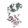
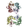
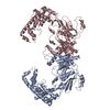
| Title | Dual specific bH1 Fab in complex with the extracellular domain of HER2/ErbB-2 | ||||||
 Components Components |
| ||||||
 Keywords Keywords |  TRANSFERASE / Fab Complex / ATP-binding / Glycoprotein / Kinase / Membrane / Nucleotide-binding / Phosphorylation / Receptor / Transmembrane / Tyrosine-protein kinase TRANSFERASE / Fab Complex / ATP-binding / Glycoprotein / Kinase / Membrane / Nucleotide-binding / Phosphorylation / Receptor / Transmembrane / Tyrosine-protein kinase | ||||||
| Function / homology |  Function and homology information Function and homology informationnegative regulation of immature T cell proliferation in thymus / ERBB3:ERBB2 complex / ERBB2-ERBB4 signaling pathway / GRB7 events in ERBB2 signaling / immature T cell proliferation in thymus / RNA polymerase I core binding / semaphorin receptor complex / regulation of microtubule-based process / ErbB-3 class receptor binding / Sema4D induced cell migration and growth-cone collapse ...negative regulation of immature T cell proliferation in thymus / ERBB3:ERBB2 complex / ERBB2-ERBB4 signaling pathway / GRB7 events in ERBB2 signaling / immature T cell proliferation in thymus / RNA polymerase I core binding / semaphorin receptor complex / regulation of microtubule-based process / ErbB-3 class receptor binding / Sema4D induced cell migration and growth-cone collapse / motor neuron axon guidance / neurotransmitter receptor localization to postsynaptic specialization membrane / PLCG1 events in ERBB2 signaling / ERBB2-EGFR signaling pathway / positive regulation of Rho protein signal transduction / ERBB2 Activates PTK6 Signaling / neuromuscular junction development / Drug-mediated inhibition of ERBB2 signaling / Resistance of ERBB2 KD mutants to trastuzumab / Resistance of ERBB2 KD mutants to sapitinib / Resistance of ERBB2 KD mutants to tesevatinib / Resistance of ERBB2 KD mutants to neratinib / Resistance of ERBB2 KD mutants to osimertinib / Resistance of ERBB2 KD mutants to afatinib / Resistance of ERBB2 KD mutants to AEE788 / Resistance of ERBB2 KD mutants to lapatinib / Drug resistance in ERBB2 TMD/JMD mutants / enzyme-linked receptor protein signaling pathway / ERBB2-ERBB3 signaling pathway / positive regulation of transcription by RNA polymerase I / oligodendrocyte differentiation / ERBB2 Regulates Cell Motility / semaphorin-plexin signaling pathway / PI3K events in ERBB2 signaling / positive regulation of protein targeting to membrane / positive regulation of cell adhesion / regulation of angiogenesis / coreceptor activity / Schwann cell development / Signaling by ERBB2 / cellular response to epidermal growth factor stimulus / myelination / TFAP2 (AP-2) family regulates transcription of growth factors and their receptors / GRB2 events in ERBB2 signaling / Downregulation of ERBB2:ERBB3 signaling / transmembrane receptor protein tyrosine kinase activity / neurogenesis / SHC1 events in ERBB2 signaling / Constitutive Signaling by Overexpressed ERBB2 / basal plasma membrane / regulation of ERK1 and ERK2 cascade / positive regulation of translation / phosphatidylinositol 3-kinase/protein kinase B signal transduction / positive regulation of epithelial cell proliferation / Signaling by ERBB2 TMD/JMD mutants / neuromuscular junction / positive regulation of MAP kinase activity / wound healing / Signaling by ERBB2 ECD mutants / Signaling by ERBB2 KD Mutants / receptor protein-tyrosine kinase / neuron differentiation / receptor tyrosine kinase binding / cellular response to growth factor stimulus / Downregulation of ERBB2 signaling / ruffle membrane / peptidyl-tyrosine phosphorylation / cell surface receptor protein tyrosine kinase signaling pathway / Constitutive Signaling by Aberrant PI3K in Cancer / transmembrane signaling receptor activity / PIP3 activates AKT signaling / presynaptic membrane / myelin sheath / heart development / PI5P, PP2A and IER3 Regulate PI3K/AKT Signaling / RAF/MAP kinase cascade / positive regulation of cell growth / basolateral plasma membrane / protein tyrosine kinase activity / positive regulation of MAPK cascade / cell surface receptor signaling pathway / receptor complex / early endosome / endosome membrane / intracellular signal transduction / positive regulation of protein phosphorylation / apical plasma membrane / protein heterodimerization activity / protein phosphorylation / signaling receptor binding / positive regulation of cell population proliferation / negative regulation of apoptotic process / perinuclear region of cytoplasm / signal transduction / nucleoplasm / ATP binding / membrane / identical protein binding / nucleus / plasma membraneSimilarity search - Function | ||||||
| Biological species |  Homo sapiens (human) Homo sapiens (human) | ||||||
| Method | X-RAY DIFFRACTION / SYNCHROTRON / MOLECULAR REPLACEMENT / molecular replacement / Resolution: 2.9 Å | ||||||
 Authors Authors | Bostrom, J.M. / Wiesmann, C. / Appleton, B.A. | ||||||
 Citation Citation | Journal: Science / Year: 2009 Title: Variants of the antibody herceptin that interact with HER2 and VEGF at the antigen binding site Authors: Bostrom, J. / Yu, S.F. / Kan, D. / Appleton, B.A. / Lee, C.V. / Billeci, K. / Man, W. / Peale, F. / Ross, S. / Wiesmann, C. / Fuh, G. | ||||||
| History |
|
- Structure visualization
| Structure viewer | Molecule: MolmilJmol/JSmol |
|---|
- Downloads & links
-Download
| PDBx/mmCIF format | 3be1.cif.gz | 210.3 KB | Display | PDBx/mmCIF format |
|---|---|---|---|---|
| PDB format | pdb3be1.ent.gz | 165.7 KB | Display | PDB format |
| PDBx/mmJSON format | 3be1.json.gz | Tree view | PDBx/mmJSON format | |
| Others |  Other downloads Other downloads |
-Validation report
| Arichive directory | https://data.pdbj.org/pub/pdb/validation_reports/be/3be1ftp://data.pdbj.org/pub/pdb/validation_reports/be/3be1 | HTTPS FTP |
|---|
-Related structure data
| Related structure data | 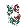 3bdySC S: Starting model for refinement C: citing same article ( |
|---|---|
| Similar structure data |


-Links
 PDBj
PDBj


- Assembly
| Deposited unit | 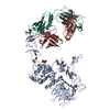
| ||||||||
|---|---|---|---|---|---|---|---|---|---|
| 1 |
| ||||||||
| Unit cell |
|
-Components
| #1: Protein | Mass: 68794.086 Da / Num. of mol.: 1 / Fragment: sequence database residues 23-646 Source method: isolated from a genetically manipulated source Source: (gene. exp.) Homo sapiens (human) / Gene: ERBB2, HER2, NEU, NGL / Cell line (production host): Chinese Hamster Ovary / Production host:   Cricetulus griseus (Chinese hamster) Cricetulus griseus (Chinese hamster)References: UniProt: P04626, receptor protein-tyrosine kinase | ||
|---|---|---|---|
| #2: Antibody | Mass: 24557.451 Da / Num. of mol.: 1 Source method: isolated from a genetically manipulated source Source: (gene. exp.) Homo sapiens (human) / Production host:  Escherichia coli (E. coli) / References: PDB-3BDY Escherichia coli (E. coli) / References: PDB-3BDY | ||
| #3: Antibody | Mass: 23956.588 Da / Num. of mol.: 1 Source method: isolated from a genetically manipulated source Source: (gene. exp.) Homo sapiens (human) / Production host: Escherichia coli (E. coli) / References: PDB-3BDY | ||
| #4: Sugar | N-Acetylglucosamine  Type: D-saccharide, beta linking / Mass: 221.208 Da / Num. of mol.: 2 Type: D-saccharide, beta linking / Mass: 221.208 Da / Num. of mol.: 2Source method: isolated from a genetically manipulated source Formula: C8H15NO6 #5: Chemical | ChemComp-MES / | MES (buffer)  Mass: 195.237 Da / Num. of mol.: 1 / Source method: obtained synthetically / Formula: C6H13NO4S / Comment: pH buffer*YM Mass: 195.237 Da / Num. of mol.: 1 / Source method: obtained synthetically / Formula: C6H13NO4S / Comment: pH buffer*YM |
-Experimental details
-Experiment
| Experiment | Method: X-RAY DIFFRACTION / Number of used crystals: 1 |
|---|
- Sample preparation
| Crystal | Density Matthews: 3.18 Å3/Da / Density % sol: 61.34 % |
|---|---|
| Crystal grow | Temperature: 292 K / Method: vapor diffusion, hanging drop / pH: 8 Details: Crystals of the Fab/HER2 complex were obtained by mixing protein solution (11 mg/ml protein, 25 mM Tris-HCl pH 8 and 150 mM sodium chloride) with crystallization buffer containing 25% w/v ...Details: Crystals of the Fab/HER2 complex were obtained by mixing protein solution (11 mg/ml protein, 25 mM Tris-HCl pH 8 and 150 mM sodium chloride) with crystallization buffer containing 25% w/v PEG2000, 0.1M MES pH 6.5. Before data collection the crystals were flash frozen in liquid nitrogen with 20% Ethylene Glycol as cryo-protectant, pH 8.0, VAPOR DIFFUSION, HANGING DROP, temperature 292K |
-Data collection
| Diffraction | Mean temperature: 100 K |
|---|---|
| Diffraction source | Source: SYNCHROTRON / Site: ALS  / Beamline: 5.0.2 / Wavelength: 1 Å / Beamline: 5.0.2 / Wavelength: 1 Å |
| Detector | Type: ADSC QUANTUM 315 / Detector: CCD / Date: Jul 13, 2006 |
| Radiation | Protocol: SINGLE WAVELENGTH / Monochromatic (M) / Laue (L): M / Scattering type: x-ray |
| Radiation wavelength | Wavelength: 1 Å / Relative weight: 1 |
| Reflection | Resolution: 2.9→50 Å / Num. all: 34149 / Num. obs: 34149 / % possible obs: 100 % / Redundancy: 5.7 % / Rsym value: 0.095 / Χ2: 0.981 / Net I/σ(I): 18.5 |
| Reflection shell | Resolution: 2.9→3 Å / Redundancy: 5.8 % / Num. unique all: 3328 / Rsym value: 0.658 / Χ2: 0.895 / % possible all: 100 |
-Phasing
| Phasing | Method: molecular replacement |
|---|
- Processing
| Software |
| |||||||||||||||||||||||||||||||||||||||||||||||||||||||||||||||||||||||||||||||||||||||||||||||||||||||||||||||||||||||||||||||||||||||||||||||||||||||||||||||||||||||||||||||||||||||||||||||||||||||||||||||||||||||||||||||||
|---|---|---|---|---|---|---|---|---|---|---|---|---|---|---|---|---|---|---|---|---|---|---|---|---|---|---|---|---|---|---|---|---|---|---|---|---|---|---|---|---|---|---|---|---|---|---|---|---|---|---|---|---|---|---|---|---|---|---|---|---|---|---|---|---|---|---|---|---|---|---|---|---|---|---|---|---|---|---|---|---|---|---|---|---|---|---|---|---|---|---|---|---|---|---|---|---|---|---|---|---|---|---|---|---|---|---|---|---|---|---|---|---|---|---|---|---|---|---|---|---|---|---|---|---|---|---|---|---|---|---|---|---|---|---|---|---|---|---|---|---|---|---|---|---|---|---|---|---|---|---|---|---|---|---|---|---|---|---|---|---|---|---|---|---|---|---|---|---|---|---|---|---|---|---|---|---|---|---|---|---|---|---|---|---|---|---|---|---|---|---|---|---|---|---|---|---|---|---|---|---|---|---|---|---|---|---|---|---|---|---|---|---|---|---|---|---|---|---|---|---|---|---|---|---|---|---|
| Refinement | Method to determine structure: MOLECULAR REPLACEMENT Starting model: PDB entry 3BDY Resolution: 2.9→30 Å / Cor.coef. Fo:Fc: 0.927 / Cor.coef. Fo:Fc free: 0.886 / SU B: 34.265 / SU ML: 0.304 / TLS residual ADP flag: LIKELY RESIDUAL / Cross valid method: THROUGHOUT / σ(F): 0 / ESU R: 1.776 / ESU R Free: 0.407 / Stereochemistry target values: MAXIMUM LIKELIHOOD / Details: HYDROGENS HAVE BEEN ADDED IN THE RIDING POSITIONS
| |||||||||||||||||||||||||||||||||||||||||||||||||||||||||||||||||||||||||||||||||||||||||||||||||||||||||||||||||||||||||||||||||||||||||||||||||||||||||||||||||||||||||||||||||||||||||||||||||||||||||||||||||||||||||||||||||
| Solvent computation | Ion probe radii: 0.8 Å / Shrinkage radii: 0.8 Å / VDW probe radii: 1.4 Å / Solvent model: MASK | |||||||||||||||||||||||||||||||||||||||||||||||||||||||||||||||||||||||||||||||||||||||||||||||||||||||||||||||||||||||||||||||||||||||||||||||||||||||||||||||||||||||||||||||||||||||||||||||||||||||||||||||||||||||||||||||||
| Displacement parameters | Biso mean: 57.135 Å2
| |||||||||||||||||||||||||||||||||||||||||||||||||||||||||||||||||||||||||||||||||||||||||||||||||||||||||||||||||||||||||||||||||||||||||||||||||||||||||||||||||||||||||||||||||||||||||||||||||||||||||||||||||||||||||||||||||
| Refinement step | Cycle: LAST / Resolution: 2.9→30 Å
| |||||||||||||||||||||||||||||||||||||||||||||||||||||||||||||||||||||||||||||||||||||||||||||||||||||||||||||||||||||||||||||||||||||||||||||||||||||||||||||||||||||||||||||||||||||||||||||||||||||||||||||||||||||||||||||||||
| Refine LS restraints |
| |||||||||||||||||||||||||||||||||||||||||||||||||||||||||||||||||||||||||||||||||||||||||||||||||||||||||||||||||||||||||||||||||||||||||||||||||||||||||||||||||||||||||||||||||||||||||||||||||||||||||||||||||||||||||||||||||
| LS refinement shell | Resolution: 2.9→2.959 Å / Total num. of bins used: 25
| |||||||||||||||||||||||||||||||||||||||||||||||||||||||||||||||||||||||||||||||||||||||||||||||||||||||||||||||||||||||||||||||||||||||||||||||||||||||||||||||||||||||||||||||||||||||||||||||||||||||||||||||||||||||||||||||||
| Refinement TLS params. | Method: refined / Refine-ID: X-RAY DIFFRACTION
| |||||||||||||||||||||||||||||||||||||||||||||||||||||||||||||||||||||||||||||||||||||||||||||||||||||||||||||||||||||||||||||||||||||||||||||||||||||||||||||||||||||||||||||||||||||||||||||||||||||||||||||||||||||||||||||||||
| Refinement TLS group |
|
