 Movie
Movie Controller

[English] 日本語
 Yorodumi
- PDB-5xhg: Crystal structure of Trastuzumab Fab fragment bearing Ne-(o-azido... -
+ Open data

- Basic information
| Entry | Database: PDB / ID: 5xhg | ||||||
|---|---|---|---|---|---|---|---|
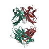
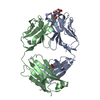
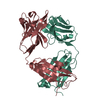
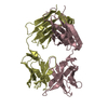
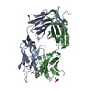
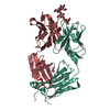
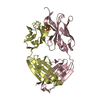
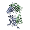
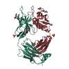
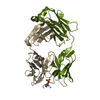
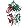
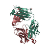
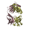
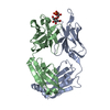
| Title | Crystal structure of Trastuzumab Fab fragment bearing Ne-(o-azidobenzyloxycarbonyl)-L-lysine | ||||||
 Components Components |
| ||||||
 Keywords Keywords |  IMMUNE SYSTEM / FAB IMMUNE SYSTEM / FAB | ||||||
| Function / homology | Immunoglobulins / Immunoglobulin-like / Sandwich / Mainly Beta / (2-azidophenyl)methyl hydrogen carbonate / DI(HYDROXYETHYL)ETHER Function and homology information Function and homology information | ||||||
| Biological species |  MUS MUSCULUS (house mouse) MUS MUSCULUS (house mouse) | ||||||
| Method | X-RAY DIFFRACTION / SYNCHROTRON / Resolution: 1.76 Å | ||||||
 Authors Authors | Kuratani, M. / Yanagisawa, T. / Sakamoto, K. / Yokoyama, S. | ||||||
 Citation Citation | Journal: Bioconjug. Chem. / Year: 2017 Title: Extensive Survey of Antibody Invariant Positions for Efficient Chemical Conjugation Using Expanded Genetic Codes. Authors: Kato, A. / Kuratani, M. / Yanagisawa, T. / Ohtake, K. / Hayashi, A. / Amano, Y. / Kimura, K. / Yokoyama, S. / Sakamoto, K. / Shiraishi, Y. | ||||||
| History |
|
- Structure visualization
| Structure viewer | Molecule: MolmilJmol/JSmol |
|---|
- Downloads & links
-Download
| PDBx/mmCIF format | 5xhg.cif.gz | 361 KB | Display | PDBx/mmCIF format |
|---|---|---|---|---|
| PDB format | pdb5xhg.ent.gz | 305.5 KB | Display | PDB format |
| PDBx/mmJSON format | 5xhg.json.gz | Tree view | PDBx/mmJSON format | |
| Others |  Other downloads Other downloads |
-Validation report
| Arichive directory | https://data.pdbj.org/pub/pdb/validation_reports/xh/5xhgftp://data.pdbj.org/pub/pdb/validation_reports/xh/5xhg | HTTPS FTP |
|---|
-Related structure data


-Links
 PDBj
PDBj

- Assembly
| Deposited unit | 
| ||||||||
|---|---|---|---|---|---|---|---|---|---|
| 1 | 
| ||||||||
| 2 | 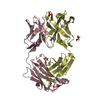
| ||||||||
| Unit cell |
|
-Components
-Antibody , 2 types, 4 molecules ACBD
| #1: Antibody | Mass: 23467.082 Da / Num. of mol.: 2 Source method: isolated from a genetically manipulated source Source: (gene. exp.) MUS MUSCULUS (house mouse) / Description: humanized mouseProduction host:  Escherichia coli str. K-12 substr. W3110 (bacteria) Escherichia coli str. K-12 substr. W3110 (bacteria)#2: Antibody | Mass: 23425.180 Da / Num. of mol.: 2 Source method: isolated from a genetically manipulated source Source: (gene. exp.) MUS MUSCULUS (house mouse) / Description: humanized mouseProduction host: Escherichia coli str. K-12 substr. W3110 (bacteria) |
|---|
-Non-polymers , 5 types, 1275 molecules 


| #3: Chemical | ChemComp-OAZ / ( Mass: 193.159 Da / Num. of mol.: 1 / Source method: obtained synthetically / Formula: C8H7N3O2 Mass: 193.159 Da / Num. of mol.: 1 / Source method: obtained synthetically / Formula: C8H7N3O2 | ||||||
|---|---|---|---|---|---|---|---|
| #4: Chemical | Sulfate Mass: 96.063 Da / Num. of mol.: 3 / Source method: obtained synthetically / Formula: SO4 Mass: 96.063 Da / Num. of mol.: 3 / Source method: obtained synthetically / Formula: SO4#5: Chemical | ChemComp-EDO / Ethylene glycol Mass: 62.068 Da / Num. of mol.: 9 / Source method: obtained synthetically / Formula: C2H6O2 Mass: 62.068 Da / Num. of mol.: 9 / Source method: obtained synthetically / Formula: C2H6O2#6: Chemical | Diethylene glycol Mass: 106.120 Da / Num. of mol.: 2 / Source method: obtained synthetically / Formula: C4H10O3 Mass: 106.120 Da / Num. of mol.: 2 / Source method: obtained synthetically / Formula: C4H10O3#7: Water | ChemComp-HOH / | WaterMass: 18.015 Da / Num. of mol.: 1260 / Source method: isolated from a natural source / Formula: H2O |
-Experimental details
-Experiment
| Experiment | Method: X-RAY DIFFRACTION / Number of used crystals: 1 |
|---|
- Sample preparation
| Crystal | Density Matthews: 2.5 Å3/Da / Density % sol: 50.77 % |
|---|---|
| Crystal grow | Temperature: 293 K / Method: vapor diffusion, sitting drop / Details: Ammonium sulfate, PEG 3350, Bis-Tris |
-Data collection
| Diffraction | Mean temperature: 100 K |
|---|---|
| Diffraction source | Source: SYNCHROTRON / Site: NSRRC  / Beamline: BL15A1 / Wavelength: 1 Å / Beamline: BL15A1 / Wavelength: 1 Å |
| Detector | Type: RAYONIX MX300HE / Detector: CCD / Date: Aug 4, 2016 |
| Radiation | Protocol: SINGLE WAVELENGTH / Monochromatic (M) / Laue (L): M / Scattering type: x-ray |
| Radiation wavelength | Wavelength: 1 Å / Relative weight: 1 |
| Reflection | Resolution: 1.76→30 Å / Num. obs: 230256 / % possible obs: 95.8 % / Redundancy: 1.3 % / Net I/σ(I): 7.92 |
- Processing
| Software |
| |||||||||||||||||||||||||||||||||||||||||||||||||||||||||||||||||||||||||||||
|---|---|---|---|---|---|---|---|---|---|---|---|---|---|---|---|---|---|---|---|---|---|---|---|---|---|---|---|---|---|---|---|---|---|---|---|---|---|---|---|---|---|---|---|---|---|---|---|---|---|---|---|---|---|---|---|---|---|---|---|---|---|---|---|---|---|---|---|---|---|---|---|---|---|---|---|---|---|---|
| Refinement | Resolution: 1.76→25.438 Å / SU ML: 0.19 / Cross valid method: FREE R-VALUE / σ(F): 0.09 / Phase error: 20.73 / Stereochemistry target values: ML
| |||||||||||||||||||||||||||||||||||||||||||||||||||||||||||||||||||||||||||||
| Solvent computation | Shrinkage radii: 0.9 Å / VDW probe radii: 1.11 Å / Solvent model: FLAT BULK SOLVENT MODEL | |||||||||||||||||||||||||||||||||||||||||||||||||||||||||||||||||||||||||||||
| Refinement step | Cycle: LAST / Resolution: 1.76→25.438 Å
| |||||||||||||||||||||||||||||||||||||||||||||||||||||||||||||||||||||||||||||
| Refine LS restraints |
| |||||||||||||||||||||||||||||||||||||||||||||||||||||||||||||||||||||||||||||
| LS refinement shell |
| |||||||||||||||||||||||||||||||||||||||||||||||||||||||||||||||||||||||||||||
| Refinement TLS params. | Method: refined / Origin x: 21.188 Å / Origin y: 63.57 Å / Origin z: 7.8992 Å
| |||||||||||||||||||||||||||||||||||||||||||||||||||||||||||||||||||||||||||||
| Refinement TLS group | Selection details: all |
