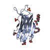
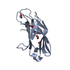
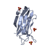
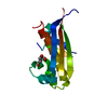
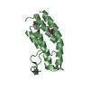
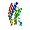
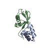
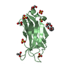
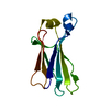
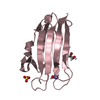
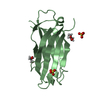
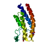
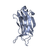
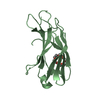
登録情報 データベース : PDB / ID : 6btyタイトル Crystal structure of the PI3KC2alpha C2 domain in space group P41212 Phosphatidylinositol 4-phosphate 3-kinase C2 domain-containing subunit alpha キーワード / / / / 機能・相同性 分子機能 ドメイン・相同性 構成要素
/ / / / / / / / / / / / / / / / / / / / / / / / / / / / / / / / / / / / / / / / / / / / / / / / / / / / / / / / / / / / / / / / / / / / / / / / / / / / / / / / / / / / / / / / / / / / / / / / / / / 生物種 Homo sapiens (ヒト)手法 / / / 解像度 : 1.678 Å データ登録者 Chen, K.-E. / Collins, B.M. ジャーナル : Structure / 年 : 2018タイトル : Molecular Basis for Membrane Recruitment by the PX and C2 Domains of Class II Phosphoinositide 3-Kinase-C2α.著者 : Kai-En Chen / Vikas A Tillu / Mintu Chandra / Brett M Collins / 要旨 : Phosphorylation of phosphoinositides by the class II phosphatidylinositol 3-kinase (PI3K) PI3K-C2α is essential for many processes, including neuroexocytosis and formation of clathrin-coated ... Phosphorylation of phosphoinositides by the class II phosphatidylinositol 3-kinase (PI3K) PI3K-C2α is essential for many processes, including neuroexocytosis and formation of clathrin-coated vesicles. A defining feature of the class II PI3Ks is a C-terminal module composed of phox-homology (PX) and C2 membrane interacting domains; however, the mechanisms that control their specific cellular localization remain poorly understood. Here we report the crystal structure of the C2 domain of PI3K-C2α in complex with the phosphoinositide head-group mimic inositol hexaphosphate, revealing two distinct pockets for membrane binding. The C2 domain preferentially binds to phosphatidylinositol 4,5-bisphosphate and phosphatidylinositol (3,4,5)-trisphosphate, and low-resolution structures of the combined PX-C2 module by small-angle X-ray scattering reveal a compact conformation in which cooperative lipid binding by each domain binding can occur. Finally, we demonstrate an unexpected role for calcium in perturbing the membrane interactions of the PX-C2 module, which we speculate may be important for regulating the activity of PI3K-C2α. 履歴 登録 2017年12月8日 登録サイト / 処理サイト 改定 1.0 2018年10月17日 Provider / タイプ 改定 1.1 2018年12月19日 Group / Database references / カテゴリ / Item / _citation.page_first改定 1.2 2023年10月4日 Group Advisory / Data collection ... Advisory / Data collection / Database references / Refinement description カテゴリ chem_comp_atom / chem_comp_bond ... chem_comp_atom / chem_comp_bond / database_2 / pdbx_initial_refinement_model / pdbx_unobs_or_zero_occ_atoms Item / _database_2.pdbx_database_accession
すべて表示 表示を減らす
 ムービー
ムービー コントローラー


 データを開く
 基本情報
基本情報 要素
要素 キーワード
キーワード TRANSFERASE (転移酵素) /
  機能・相同性情報
 データ登録者
 引用
引用
 構造の表示
構造の表示 ダウンロードとリンク
 その他のダウンロード


 PDBj
PDBj


 集合体
集合体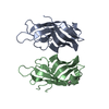


 分子量: 264.315 Da / 分子数: 1 / 由来タイプ: 合成 / 式: C12H24O6
 分子量: 18.015 Da / 分子数: 184 / 由来タイプ: 天然 / 式: H2O
 試料調製
試料調製 解析
解析