 Movie
Movie Controller

[English] 日本語
 Yorodumi
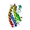
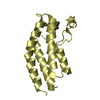
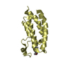
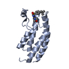
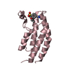
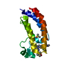
- PDB-6bu0: Crystal structure of the PI3KC2alpha C2 domain in complex with IP6 -
+ Open data

- Basic information
| Entry | Database: PDB / ID: 6bu0 | ||||||
|---|---|---|---|---|---|---|---|
| Title | Crystal structure of the PI3KC2alpha C2 domain in complex with IP6 | ||||||
 Components Components | Phosphatidylinositol 4-phosphate 3-kinase C2 domain-containing subunit alpha | ||||||
 Keywords Keywords |  TRANSFERASE / C2 domain / lipid binding / phosphoinositide / PI3-kinase TRANSFERASE / C2 domain / lipid binding / phosphoinositide / PI3-kinase | ||||||
| Function / homology |  Function and homology information Function and homology informationvascular associated smooth muscle contraction / Synthesis of PIPs at the late endosome membrane / Synthesis of PIPs at the early endosome membrane / Synthesis of PIPs at the Golgi membrane / phosphatidylinositol-4-phosphate 3-kinase / clathrin coat assembly / membrane organization / phosphatidylinositol biosynthetic process / phosphatidylinositol 3-kinase complex / 1-phosphatidylinositol-4-phosphate 3-kinase activity ...vascular associated smooth muscle contraction / Synthesis of PIPs at the late endosome membrane / Synthesis of PIPs at the early endosome membrane / Synthesis of PIPs at the Golgi membrane / phosphatidylinositol-4-phosphate 3-kinase / clathrin coat assembly / membrane organization / phosphatidylinositol biosynthetic process / phosphatidylinositol 3-kinase complex / 1-phosphatidylinositol-4-phosphate 3-kinase activity / 1-phosphatidylinositol-4,5-bisphosphate 3-kinase activity / phosphatidylinositol-4,5-bisphosphate 3-kinase / phosphatidylinositol 3-kinase / clathrin-coated vesicle / phosphatidylinositol-3-phosphate biosynthetic process / 1-phosphatidylinositol-3-kinase activity / clathrin binding / positive regulation of cell migration involved in sprouting angiogenesis / Golgi Associated Vesicle Biogenesis / exocytosis / Synthesis of PIPs at the plasma membrane / platelet-derived growth factor receptor signaling pathway / positive regulation of autophagy / phosphatidylinositol binding / phosphatidylinositol 3-kinase/protein kinase B signal transduction / epidermal growth factor receptor signaling pathway / trans-Golgi network / endocytosis / insulin receptor signaling pathway / Clathrin-mediated endocytosis / vesicle / phosphorylation / intracellular membrane-bounded organelle / extracellular exosome / nucleoplasm / ATP binding / membrane / plasma membrane / cytosol / cytoplasmSimilarity search - Function | ||||||
| Biological species |  Homo sapiens (human) Homo sapiens (human) | ||||||
| Method | X-RAY DIFFRACTION / SYNCHROTRON / MOLECULAR REPLACEMENT / molecular replacement / Resolution: 2.427 Å | ||||||
 Authors Authors | Chen, K.-E. / Collins, B.M. | ||||||
 Citation Citation | Journal: Structure / Year: 2018 Title: Molecular Basis for Membrane Recruitment by the PX and C2 Domains of Class II Phosphoinositide 3-Kinase-C2α. Authors: Kai-En Chen / Vikas A Tillu / Mintu Chandra / Brett M Collins /  Abstract: Phosphorylation of phosphoinositides by the class II phosphatidylinositol 3-kinase (PI3K) PI3K-C2α is essential for many processes, including neuroexocytosis and formation of clathrin-coated ...Phosphorylation of phosphoinositides by the class II phosphatidylinositol 3-kinase (PI3K) PI3K-C2α is essential for many processes, including neuroexocytosis and formation of clathrin-coated vesicles. A defining feature of the class II PI3Ks is a C-terminal module composed of phox-homology (PX) and C2 membrane interacting domains; however, the mechanisms that control their specific cellular localization remain poorly understood. Here we report the crystal structure of the C2 domain of PI3K-C2α in complex with the phosphoinositide head-group mimic inositol hexaphosphate, revealing two distinct pockets for membrane binding. The C2 domain preferentially binds to phosphatidylinositol 4,5-bisphosphate and phosphatidylinositol (3,4,5)-trisphosphate, and low-resolution structures of the combined PX-C2 module by small-angle X-ray scattering reveal a compact conformation in which cooperative lipid binding by each domain binding can occur. Finally, we demonstrate an unexpected role for calcium in perturbing the membrane interactions of the PX-C2 module, which we speculate may be important for regulating the activity of PI3K-C2α. | ||||||
| History |
|
- Structure visualization
| Structure viewer | Molecule: MolmilJmol/JSmol |
|---|
- Downloads & links
-Download
| PDBx/mmCIF format | 6bu0.cif.gz | 95 KB | Display | PDBx/mmCIF format |
|---|---|---|---|---|
| PDB format | pdb6bu0.ent.gz | 72 KB | Display | PDB format |
| PDBx/mmJSON format | 6bu0.json.gz | Tree view | PDBx/mmJSON format | |
| Others |  Other downloads Other downloads |
-Validation report
| Arichive directory | https://data.pdbj.org/pub/pdb/validation_reports/bu/6bu0ftp://data.pdbj.org/pub/pdb/validation_reports/bu/6bu0 | HTTPS FTP |
|---|
-Related structure data
| Related structure data | 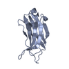 6btyC 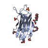 6btzC  6bubC 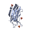 2b3rS S: Starting model for refinement C: citing same article ( |
|---|---|
| Similar structure data |

-Links
 PDBj
PDBj


- Assembly
| Deposited unit | 
| ||||||||
|---|---|---|---|---|---|---|---|---|---|
| 1 | 
| ||||||||
| 2 | 
| ||||||||
| 3 | 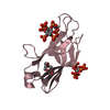
| ||||||||
| Unit cell |
|
-Components
| #1: Protein | Mass: 14872.116 Da / Num. of mol.: 3 / Fragment: C2 domain / Mutation: E1654G Source method: isolated from a genetically manipulated source Source: (gene. exp.) Homo sapiens (human) / Gene: PIK3C2A / Production host:  Escherichia coli (E. coli) / Strain (production host): BL21(DE3) Escherichia coli (E. coli) / Strain (production host): BL21(DE3)References: UniProt: O00443, phosphatidylinositol-4-phosphate 3-kinase#2: Chemical | ChemComp-FMT / Formic acid  Mass: 46.025 Da / Num. of mol.: 5 / Source method: obtained synthetically / Formula: CH2O2 Mass: 46.025 Da / Num. of mol.: 5 / Source method: obtained synthetically / Formula: CH2O2#3: Chemical | ChemComp-IHP / Phytic acid  Mass: 660.035 Da / Num. of mol.: 4 / Source method: obtained synthetically / Formula: C6H18O24P6 Mass: 660.035 Da / Num. of mol.: 4 / Source method: obtained synthetically / Formula: C6H18O24P6#4: Chemical | ChemComp-O4B / | 18-Crown-6  Mass: 264.315 Da / Num. of mol.: 1 / Source method: obtained synthetically / Formula: C12H24O6 Mass: 264.315 Da / Num. of mol.: 1 / Source method: obtained synthetically / Formula: C12H24O6#5: Water | ChemComp-HOH / | Water Mass: 18.015 Da / Num. of mol.: 23 / Source method: isolated from a natural source / Formula: H2O Mass: 18.015 Da / Num. of mol.: 23 / Source method: isolated from a natural source / Formula: H2O |
|---|
-Experimental details
-Experiment
| Experiment | Method: X-RAY DIFFRACTION / Number of used crystals: 1 |
|---|
- Sample preparation
| Crystal | Density Matthews: 2.52 Å3/Da / Density % sol: 51.1 % / Mosaicity: 0.11 ° |
|---|---|
| Crystal grow | Temperature: 295 K / Method: vapor diffusion, hanging drop / pH: 7.5 / Details: PEG 8000 |
-Data collection
| Diffraction | Mean temperature: 100 K |
|---|---|
| Diffraction source | Source: SYNCHROTRON / Site: Australian Synchrotron / Beamline: MX2 / Wavelength: 1 Å |
| Detector | Type: ADSC QUANTUM 315r / Detector: CCD / Date: Apr 19, 2017 |
| Radiation | Protocol: SINGLE WAVELENGTH / Monochromatic (M) / Laue (L): M / Scattering type: x-ray |
| Radiation wavelength | Wavelength: 1 Å / Relative weight: 1 |
| Reflection | Resolution: 2.43→45.83 Å / Num. obs: 17713 / % possible obs: 99.6 % / Redundancy: 6.5 % / Biso Wilson estimate: 56.8 Å2 / CC1/2: 0.999 / Rmerge(I) obs: 0.046 / Rpim(I) all: 0.02 / Rrim(I) all: 0.051 / Net I/σ(I): 16.8 / Num. measured all: 114498 |
| Reflection shell | Resolution: 2.43→2.52 Å / Redundancy: 6.6 % / Rmerge(I) obs: 0.496 / Num. measured all: 11479 / Num. unique obs: 1747 / CC1/2: 0.915 / Rpim(I) all: 0.208 / Rrim(I) all: 0.538 / Net I/σ(I) obs: 2.7 / % possible all: 97 |
-Phasing
| Phasing | Method: molecular replacement | |||||||||
|---|---|---|---|---|---|---|---|---|---|---|
| Phasing MR | Model details: Phaser MODE: MR_AUTO
|
- Processing
| Software |
| |||||||||||||||||||||||||||||||||||||||||||||||||
|---|---|---|---|---|---|---|---|---|---|---|---|---|---|---|---|---|---|---|---|---|---|---|---|---|---|---|---|---|---|---|---|---|---|---|---|---|---|---|---|---|---|---|---|---|---|---|---|---|---|---|
| Refinement | Method to determine structure: MOLECULAR REPLACEMENT Starting model: 2B3R Resolution: 2.427→43.094 Å / SU ML: 0.32 / Cross valid method: THROUGHOUT / σ(F): 1.38 / Phase error: 27.99 / Stereochemistry target values: MLHL
| |||||||||||||||||||||||||||||||||||||||||||||||||
| Solvent computation | Shrinkage radii: 0.9 Å / VDW probe radii: 1.11 Å / Solvent model: FLAT BULK SOLVENT MODEL | |||||||||||||||||||||||||||||||||||||||||||||||||
| Displacement parameters | Biso max: 152.43 Å2 / Biso mean: 58.7087 Å2 / Biso min: 30 Å2 | |||||||||||||||||||||||||||||||||||||||||||||||||
| Refinement step | Cycle: final / Resolution: 2.427→43.094 Å
| |||||||||||||||||||||||||||||||||||||||||||||||||
| Refine LS restraints |
| |||||||||||||||||||||||||||||||||||||||||||||||||
| LS refinement shell | Refine-ID: X-RAY DIFFRACTION / Rfactor Rfree error: 0 / Total num. of bins used: 6
|
