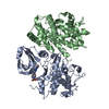
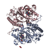
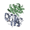
Entry Database : PDB / ID : 6athTitle Cdk2/cyclin A/p27-KID-deltaC Cyclin-A2 Cyclin-dependent kinase 2 Cyclin-dependent kinase inhibitor 1B Keywords / / / / / / / Function / homology Function Domain/homology Component
/ / / / / / / / / / / / / / / / / / / / / / / / / / / / / / / / / / / / / / / / / / / / / / / / / / / / / / / / / / / / / / / / / / / / / / / / / / / / / / / / / / / / / / / / / / / / / / / / / / / / / / / / / / / / / / / / / / / / / / / / / / / / / / / / / / / / / / / / / / / / / / / / / / / / Biological species Homo sapiens (human)Method / / / Resolution : 1.82 Å Authors White, S.W. / Yun, M. Funding support Organization Grant number Country National Institutes of Health/National Cancer Institute (NIH/NCI) R01 CA82491, P30 CA21765
Journal : Nat Commun / Year : 2019Title : Dynamic anticipation by Cdk2/Cyclin A-bound p27 mediates signal integration in cell cycle regulation.Authors: Tsytlonok, M. / Sanabria, H. / Wang, Y. / Felekyan, S. / Hemmen, K. / Phillips, A.H. / Yun, M.K. / Waddell, M.B. / Park, C.G. / Vaithiyalingam, S. / Iconaru, L. / White, S.W. / Tompa, P. / ... Authors : Tsytlonok, M. / Sanabria, H. / Wang, Y. / Felekyan, S. / Hemmen, K. / Phillips, A.H. / Yun, M.K. / Waddell, M.B. / Park, C.G. / Vaithiyalingam, S. / Iconaru, L. / White, S.W. / Tompa, P. / Seidel, C.A.M. / Kriwacki, R. History Deposition Aug 29, 2017 Deposition site / Processing site Revision 1.0 Sep 12, 2018 Provider / Type Revision 1.1 Feb 20, 2019 Group / Data collection / Category / Item Revision 1.2 Apr 10, 2019 Group / Database references / Category Item _citation.country / _citation.journal_abbrev ... _citation.country / _citation.journal_abbrev / _citation.journal_id_CSD / _citation.journal_id_ISSN / _citation.journal_volume / _citation.pdbx_database_id_DOI / _citation.year Revision 1.3 Apr 24, 2019 Group / Database references / Category / citation_authorItem _citation.page_first / _citation.page_last ... _citation.page_first / _citation.page_last / _citation.pdbx_database_id_PubMed / _citation.title Revision 1.4 Dec 4, 2019 Group / Category / Item Revision 1.5 Oct 4, 2023 Group / Database references / Refinement descriptionCategory chem_comp_atom / chem_comp_bond ... chem_comp_atom / chem_comp_bond / database_2 / pdbx_initial_refinement_model Item / _database_2.pdbx_database_accession
Show all Show less
 Movie
Movie Controller


 Open data
 Basic information
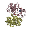
 Components
 Keywords
 TRANSFERASE / Inhibitor /
  Function and homology information
 Authors
 United States, 1items
  Citation
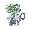
 Structure visualization
 Downloads & links
 Other downloads


 PDBj
PDBj


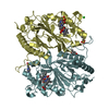
 Assembly
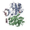


 Mass: 96.063 Da / Num. of mol.: 1 / Source method: obtained synthetically / Formula: SO4
 Mass: 18.015 Da / Num. of mol.: 399 / Source method: isolated from a natural source / Formula: H2O
 Sample preparation
 Processing