 Movie
Movie Controller

[English] 日本語
 Yorodumi
- PDB-6kmm: Crystal Structure of HEPES bound Dye Decolorizing peroxidase from... -
+ Open data

- Basic information
| Entry | Database: PDB / ID: 6kmm | ||||||
|---|---|---|---|---|---|---|---|
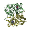
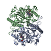
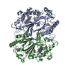
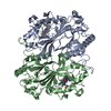
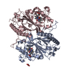
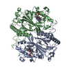
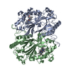
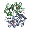
| Title | Crystal Structure of HEPES bound Dye Decolorizing peroxidase from Bacillus subtilis | ||||||
 Components Components | Deferrochelatase/peroxidase EfeB | ||||||
 Keywords Keywords |  OXIDOREDUCTASE / Dye-decolorizing peroxidase / ferredoxin like fold / HEPES OXIDOREDUCTASE / Dye-decolorizing peroxidase / ferredoxin like fold / HEPES | ||||||
| Function / homology |  Function and homology information Function and homology informationprotoporphyrin ferrochelatase / iron import into cell / ferrochelatase activity / Oxidoreductases; Acting on a peroxide as acceptor; Peroxidases / peroxidase activity / heme binding / extracellular region / metal ion binding / cytosolSimilarity search - Function | ||||||
| Biological species |  Bacillus subtilis (bacteria) Bacillus subtilis (bacteria) | ||||||
| Method | X-RAY DIFFRACTION / SYNCHROTRON / MOLECULAR REPLACEMENT / Resolution: 1.93 Å | ||||||
 Authors Authors | Dhankhar, P. / Dalal, V. / Mahto, J.K. / Kumar, P. | ||||||
| Funding support |  India, 1items India, 1items
| ||||||
 Citation Citation | Journal: Arch.Biochem.Biophys. / Year: 2020 Title: Characterization of dye-decolorizing peroxidase from Bacillus subtilis. Authors: Dhankhar, P. / Dalal, V. / Mahto, J.K. / Gurjar, B.R. / Tomar, S. / Sharma, A.K. / Kumar, P. | ||||||
| History |
|
- Structure visualization
| Structure viewer | Molecule: MolmilJmol/JSmol |
|---|
- Downloads & links
-Download
| PDBx/mmCIF format | 6kmm.cif.gz | 904.6 KB | Display | PDBx/mmCIF format |
|---|---|---|---|---|
| PDB format | pdb6kmm.ent.gz | 752.9 KB | Display | PDB format |
| PDBx/mmJSON format | 6kmm.json.gz | Tree view | PDBx/mmJSON format | |
| Others |  Other downloads Other downloads |
-Validation report
| Arichive directory | https://data.pdbj.org/pub/pdb/validation_reports/km/6kmmftp://data.pdbj.org/pub/pdb/validation_reports/km/6kmm | HTTPS FTP |
|---|
-Related structure data
| Related structure data |  6kmnC 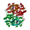 4grcS S: Starting model for refinement C: citing same article ( |
|---|---|
| Similar structure data |


-Links
 PDBj
PDBj


- Assembly
| Deposited unit | 
| ||||||||
|---|---|---|---|---|---|---|---|---|---|
| 1 | 
| ||||||||
| 2 | 
| ||||||||
| 3 | 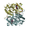
| ||||||||
| Unit cell |
|
-Components
-Protein , 1 types, 6 molecules ABCDEF
| #1: Protein | Mass: 40235.324 Da / Num. of mol.: 6 Source method: isolated from a genetically manipulated source Source: (gene. exp.) Bacillus subtilis (bacteria) / Gene: efeB, FC605_19685 / Production host: Escherichia coli BL21(DE3) (bacteria) / References: UniProt: A0A4P9FDJ4, UniProt: P39597*PLUS |
|---|
-Non-polymers , 9 types, 2126 molecules 


| #2: Chemical | ChemComp-HEM / Heme B Mass: 616.487 Da / Num. of mol.: 6 / Source method: obtained synthetically / Formula: C34H32FeN4O4 / Feature type: SUBJECT OF INVESTIGATION Mass: 616.487 Da / Num. of mol.: 6 / Source method: obtained synthetically / Formula: C34H32FeN4O4 / Feature type: SUBJECT OF INVESTIGATION#3: Chemical | ChemComp-OXY / Oxygen Mass: 31.999 Da / Num. of mol.: 6 / Source method: obtained synthetically / Formula: O2 Mass: 31.999 Da / Num. of mol.: 6 / Source method: obtained synthetically / Formula: O2#4: Chemical | ChemComp-EPE / HEPES Mass: 238.305 Da / Num. of mol.: 5 / Source method: obtained synthetically / Formula: C8H18N2O4S / Comment: pH buffer*YM Mass: 238.305 Da / Num. of mol.: 5 / Source method: obtained synthetically / Formula: C8H18N2O4S / Comment: pH buffer*YM#5: Chemical | ChemComp-GOL / Glycerol Mass: 92.094 Da / Num. of mol.: 6 / Source method: obtained synthetically / Formula: C3H8O3 Mass: 92.094 Da / Num. of mol.: 6 / Source method: obtained synthetically / Formula: C3H8O3#6: Chemical | ChemComp-NA / |  Mass: 22.990 Da / Num. of mol.: 1 / Source method: obtained synthetically / Formula: Na Mass: 22.990 Da / Num. of mol.: 1 / Source method: obtained synthetically / Formula: Na#7: Chemical | 2-Methyl-2,4-pentanediol Mass: 118.174 Da / Num. of mol.: 3 / Source method: obtained synthetically / Formula: C6H14O2 / Comment: precipitant*YM Mass: 118.174 Da / Num. of mol.: 3 / Source method: obtained synthetically / Formula: C6H14O2 / Comment: precipitant*YM#8: Chemical | ChemComp-PO4 / | Phosphate Mass: 94.971 Da / Num. of mol.: 1 / Source method: obtained synthetically / Formula: PO4 Mass: 94.971 Da / Num. of mol.: 1 / Source method: obtained synthetically / Formula: PO4#9: Chemical | Chloride Mass: 35.453 Da / Num. of mol.: 2 / Source method: obtained synthetically / Formula: Cl Mass: 35.453 Da / Num. of mol.: 2 / Source method: obtained synthetically / Formula: Cl#10: Water | ChemComp-HOH / | WaterMass: 18.015 Da / Num. of mol.: 2096 / Source method: isolated from a natural source / Formula: H2O |
|---|
-Details
| Has ligand of interest | Y |
|---|
-Experimental details
-Experiment
| Experiment | Method: X-RAY DIFFRACTION / Number of used crystals: 1 |
|---|
- Sample preparation
| Crystal | Density Matthews: 3.46 Å3/Da / Density % sol: 64.44 % |
|---|---|
| Crystal grow | Temperature: 293 K / Method: vapor diffusion, sitting drop / pH: 7 / Details: 26% MPD, 0.05 M HEPES |
-Data collection
| Diffraction | Mean temperature: 100 K / Serial crystal experiment: N |
|---|---|
| Diffraction source | Source: SYNCHROTRON / Site: ESRF  / Beamline: ID29 / Wavelength: 0.9677 Å / Beamline: ID29 / Wavelength: 0.9677 Å |
| Detector | Type: DECTRIS EIGER R 4M / Detector: PIXEL / Date: Sep 28, 2018 |
| Radiation | Monochromator: C(110) / Protocol: SINGLE WAVELENGTH / Monochromatic (M) / Laue (L): M / Scattering type: x-ray |
| Radiation wavelength | Wavelength: 0.9677 Å / Relative weight: 1 |
| Reflection | Resolution: 1.93→69.91 Å / Num. obs: 223932 / % possible obs: 92.8 % / Redundancy: 6.5 % / CC1/2: 0.99 / Net I/σ(I): 9.6 |
| Reflection shell | Resolution: 1.93→1.96 Å / Mean I/σ(I) obs: 2.2 / Num. unique obs: 10799 / CC1/2: 0.95 / % possible all: 89.4 |
- Processing
| Software |
| |||||||||||||||||||||||||||||||||||||||||||||||||||||||||||||||||||||||||||||||||||||||||||||||||||||||||||||||||||||||||||||||||||||||||||||||||||||||||||||||||||||||||||||||
|---|---|---|---|---|---|---|---|---|---|---|---|---|---|---|---|---|---|---|---|---|---|---|---|---|---|---|---|---|---|---|---|---|---|---|---|---|---|---|---|---|---|---|---|---|---|---|---|---|---|---|---|---|---|---|---|---|---|---|---|---|---|---|---|---|---|---|---|---|---|---|---|---|---|---|---|---|---|---|---|---|---|---|---|---|---|---|---|---|---|---|---|---|---|---|---|---|---|---|---|---|---|---|---|---|---|---|---|---|---|---|---|---|---|---|---|---|---|---|---|---|---|---|---|---|---|---|---|---|---|---|---|---|---|---|---|---|---|---|---|---|---|---|---|---|---|---|---|---|---|---|---|---|---|---|---|---|---|---|---|---|---|---|---|---|---|---|---|---|---|---|---|---|---|---|---|---|
| Refinement | Method to determine structure: MOLECULAR REPLACEMENT Starting model: 4GRC Resolution: 1.93→69.91 Å / Cor.coef. Fo:Fc: 0.973 / Cor.coef. Fo:Fc free: 0.959 / SU B: 8.32 / SU ML: 0.113 / Cross valid method: THROUGHOUT / σ(F): 0 / ESU R: 0.123 / ESU R Free: 0.123 / Stereochemistry target values: MAXIMUM LIKELIHOOD / Details: U VALUES : WITH TLS ADDED
| |||||||||||||||||||||||||||||||||||||||||||||||||||||||||||||||||||||||||||||||||||||||||||||||||||||||||||||||||||||||||||||||||||||||||||||||||||||||||||||||||||||||||||||||
| Solvent computation | Ion probe radii: 0.8 Å / Shrinkage radii: 0.8 Å / VDW probe radii: 1.2 Å / Solvent model: MASK | |||||||||||||||||||||||||||||||||||||||||||||||||||||||||||||||||||||||||||||||||||||||||||||||||||||||||||||||||||||||||||||||||||||||||||||||||||||||||||||||||||||||||||||||
| Displacement parameters | Biso max: 142.16 Å2 / Biso mean: 45.601 Å2 / Biso min: 22.92 Å2
| |||||||||||||||||||||||||||||||||||||||||||||||||||||||||||||||||||||||||||||||||||||||||||||||||||||||||||||||||||||||||||||||||||||||||||||||||||||||||||||||||||||||||||||||
| Refinement step | Cycle: final / Resolution: 1.93→69.91 Å
| |||||||||||||||||||||||||||||||||||||||||||||||||||||||||||||||||||||||||||||||||||||||||||||||||||||||||||||||||||||||||||||||||||||||||||||||||||||||||||||||||||||||||||||||
| Refine LS restraints |
| |||||||||||||||||||||||||||||||||||||||||||||||||||||||||||||||||||||||||||||||||||||||||||||||||||||||||||||||||||||||||||||||||||||||||||||||||||||||||||||||||||||||||||||||
| LS refinement shell | Resolution: 1.93→1.98 Å / Rfactor Rfree error: 0 / Total num. of bins used: 20
| |||||||||||||||||||||||||||||||||||||||||||||||||||||||||||||||||||||||||||||||||||||||||||||||||||||||||||||||||||||||||||||||||||||||||||||||||||||||||||||||||||||||||||||||
| Refinement TLS params. | Method: refined / Refine-ID: X-RAY DIFFRACTION
| |||||||||||||||||||||||||||||||||||||||||||||||||||||||||||||||||||||||||||||||||||||||||||||||||||||||||||||||||||||||||||||||||||||||||||||||||||||||||||||||||||||||||||||||
| Refinement TLS group |
|
