 Movie
Movie Controller

[English] 日本語
 Yorodumi
- PDB-4grc: Crystal structure of DyP-type peroxidase (SCO2276) from Streptomy... -
+ Open data

- Basic information
| Entry | Database: PDB / ID: 4grc | ||||||
|---|---|---|---|---|---|---|---|
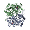
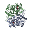
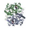
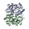
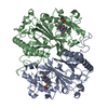
| Title | Crystal structure of DyP-type peroxidase (SCO2276) from Streptomyces coelicolor | ||||||
 Components Components | Putative membrane protein | ||||||
 Keywords Keywords | OXIDOREDUCTASE / ferridoxin-like | ||||||
| Function / homology |  Function and homology information Function and homology informationiron import into cell / ferrochelatase activity / Oxidoreductases; Acting on a peroxide as acceptor; Peroxidases / peroxidase activity / heme binding / metal ion binding / cytosolSimilarity search - Function | ||||||
| Biological species |  Streptomyces coelicolor (bacteria) Streptomyces coelicolor (bacteria) | ||||||
| Method | X-RAY DIFFRACTION / SYNCHROTRON / MOLECULAR REPLACEMENT / Resolution: 2 Å | ||||||
 Authors Authors | Lukk, T. / Hetta, A.M.A. / Jones, A. / Solbiati, J. / Majumdar, S. / Cronan, J.E. / Gerlt, J.A. / Nair, S.K. | ||||||
 Citation Citation | Journal: To be Published Title: DyP-type peroxidases from Stretptomyces and Thermobifida can modify organosolv lignin. Authors: Lukk, T. / Hetta, A.M.A. / Jones, A. / Solbiati, J. / Majumdar, S. / Cronan, J.E. / Gerlt, J.A. / Nair, S.K. | ||||||
| History |
|
- Structure visualization
| Structure viewer | Molecule: MolmilJmol/JSmol |
|---|
- Downloads & links
-Download
| PDBx/mmCIF format | 4grc.cif.gz | 97.4 KB | Display | PDBx/mmCIF format |
|---|---|---|---|---|
| PDB format | pdb4grc.ent.gz | 69.6 KB | Display | PDB format |
| PDBx/mmJSON format | 4grc.json.gz | Tree view | PDBx/mmJSON format | |
| Others |  Other downloads Other downloads |
-Validation report
| Arichive directory | https://data.pdbj.org/pub/pdb/validation_reports/gr/4grcftp://data.pdbj.org/pub/pdb/validation_reports/gr/4grc | HTTPS FTP |
|---|
-Related structure data
| Related structure data |  4gs1C  4gt2C  4gu7C  2gvkS C: citing same article ( S: Starting model for refinement |
|---|---|
| Similar structure data |


-Links
 PDBj
PDBj

- Assembly
| Deposited unit | 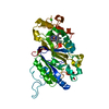
| ||||||||
|---|---|---|---|---|---|---|---|---|---|
| 1 | 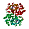
| ||||||||
| Unit cell |
| ||||||||
| Components on special symmetry positions |
|
-Components
| #1: Protein | / Dyp-type peroxidase Mass: 46750.094 Da / Num. of mol.: 1 Source method: isolated from a genetically manipulated source Source: (gene. exp.) Streptomyces coelicolor (bacteria) / Strain: ATCC BAA-471 / A3(2) / M145 / Gene: SCO2276 / Plasmid: pET-15b / Production host: Escherichia coli (E. coli) / Strain (production host): BL21(DE3) / References: UniProt: Q9RKQ2, dye decolorizing peroxidase |
|---|---|
| #2: Chemical | ChemComp-CA /   Mass: 40.078 Da / Num. of mol.: 1 / Source method: obtained synthetically / Formula: Ca Mass: 40.078 Da / Num. of mol.: 1 / Source method: obtained synthetically / Formula: Ca |
| #3: Chemical | ChemComp-HEM / Heme B  Mass: 616.487 Da / Num. of mol.: 1 / Source method: obtained synthetically / Formula: C34H32FeN4O4 Mass: 616.487 Da / Num. of mol.: 1 / Source method: obtained synthetically / Formula: C34H32FeN4O4 |
| #4: Chemical | ChemComp-PEO / Hydrogen peroxide  Mass: 34.015 Da / Num. of mol.: 1 / Source method: obtained synthetically / Formula: H2O2 Mass: 34.015 Da / Num. of mol.: 1 / Source method: obtained synthetically / Formula: H2O2 |
| #5: Water | ChemComp-HOH / Water Mass: 18.015 Da / Num. of mol.: 387 / Source method: isolated from a natural source / Formula: H2O Mass: 18.015 Da / Num. of mol.: 387 / Source method: isolated from a natural source / Formula: H2O |
-Experimental details
-Experiment
| Experiment | Method: X-RAY DIFFRACTION / Number of used crystals: 1 |
|---|
- Sample preparation
| Crystal | Density Matthews: 2.73 Å3/Da / Density % sol: 55 % |
|---|---|
| Crystal grow | Temperature: 282 K / Method: vapor diffusion, hanging drop Details: Mother liqueur contained 0.2M CaCl2, 0.1M Hepes (pH 7.5) and 28% PEG400. Protein concentration was 15 mg/mL in a buffer containing 100 mM KCl and 50 mM Hepes 8.0. 0.5 uL protein was mixed ...Details: Mother liqueur contained 0.2M CaCl2, 0.1M Hepes (pH 7.5) and 28% PEG400. Protein concentration was 15 mg/mL in a buffer containing 100 mM KCl and 50 mM Hepes 8.0. 0.5 uL protein was mixed with equal volume of mother liqueur, VAPOR DIFFUSION, HANGING DROP, temperature 282K |
-Data collection
| Diffraction | Mean temperature: 100 K | ||||||||||||||||||||||||||||||||||||||||||||||||||||||||||||||||||||||||||||||||||||||||||||||||||||||||||||||||||||||||||||||
|---|---|---|---|---|---|---|---|---|---|---|---|---|---|---|---|---|---|---|---|---|---|---|---|---|---|---|---|---|---|---|---|---|---|---|---|---|---|---|---|---|---|---|---|---|---|---|---|---|---|---|---|---|---|---|---|---|---|---|---|---|---|---|---|---|---|---|---|---|---|---|---|---|---|---|---|---|---|---|---|---|---|---|---|---|---|---|---|---|---|---|---|---|---|---|---|---|---|---|---|---|---|---|---|---|---|---|---|---|---|---|---|---|---|---|---|---|---|---|---|---|---|---|---|---|---|---|---|
| Diffraction source | Source: SYNCHROTRON / Site: APS  / Beamline: 21-ID-G / Wavelength: 0.97857 Å / Beamline: 21-ID-G / Wavelength: 0.97857 Å | ||||||||||||||||||||||||||||||||||||||||||||||||||||||||||||||||||||||||||||||||||||||||||||||||||||||||||||||||||||||||||||||
| Detector | Type: RAYONIX MX-300 / Detector: CCD / Date: Aug 22, 2010 | ||||||||||||||||||||||||||||||||||||||||||||||||||||||||||||||||||||||||||||||||||||||||||||||||||||||||||||||||||||||||||||||
| Radiation | Monochromator: C(111) / Protocol: SINGLE WAVELENGTH / Monochromatic (M) / Laue (L): M / Scattering type: x-ray | ||||||||||||||||||||||||||||||||||||||||||||||||||||||||||||||||||||||||||||||||||||||||||||||||||||||||||||||||||||||||||||||
| Radiation wavelength | Wavelength: 0.97857 Å / Relative weight: 1 | ||||||||||||||||||||||||||||||||||||||||||||||||||||||||||||||||||||||||||||||||||||||||||||||||||||||||||||||||||||||||||||||
| Reflection | Number: 643289 / Rmerge(I) obs: 0.095 / D res high: 2 Å / Num. obs: 66865 / % possible obs: 100 | ||||||||||||||||||||||||||||||||||||||||||||||||||||||||||||||||||||||||||||||||||||||||||||||||||||||||||||||||||||||||||||||
| Diffraction reflection shell |
| ||||||||||||||||||||||||||||||||||||||||||||||||||||||||||||||||||||||||||||||||||||||||||||||||||||||||||||||||||||||||||||||
| Reflection | Resolution: 2→30 Å / Num. obs: 35482 / % possible obs: 100 % / Observed criterion σ(F): 0 / Observed criterion σ(I): 0 / Redundancy: 9.6 % / Biso Wilson estimate: 23.97 Å2 / Rsym value: 0.095 / Net I/σ(I): 19.09 | ||||||||||||||||||||||||||||||||||||||||||||||||||||||||||||||||||||||||||||||||||||||||||||||||||||||||||||||||||||||||||||||
| Reflection shell |
|
- Processing
| Software |
| ||||||||||||||||||||||||||||||||||||||||||||||||||||||||||||||||||||||||||||||||||||
|---|---|---|---|---|---|---|---|---|---|---|---|---|---|---|---|---|---|---|---|---|---|---|---|---|---|---|---|---|---|---|---|---|---|---|---|---|---|---|---|---|---|---|---|---|---|---|---|---|---|---|---|---|---|---|---|---|---|---|---|---|---|---|---|---|---|---|---|---|---|---|---|---|---|---|---|---|---|---|---|---|---|---|---|---|---|
| Refinement | Method to determine structure: MOLECULAR REPLACEMENT Starting model: PDB entry 2GVK Resolution: 2→29.283 Å / Occupancy max: 1 / Occupancy min: 0.4 / FOM work R set: 0.8894 / SU ML: 0.17 / Cross valid method: thorough / σ(F): 2.02 / Phase error: 17.68 / Stereochemistry target values: ML
| ||||||||||||||||||||||||||||||||||||||||||||||||||||||||||||||||||||||||||||||||||||
| Solvent computation | Shrinkage radii: 0.9 Å / VDW probe radii: 1.11 Å / Solvent model: FLAT BULK SOLVENT MODEL | ||||||||||||||||||||||||||||||||||||||||||||||||||||||||||||||||||||||||||||||||||||
| Displacement parameters | Biso max: 76.93 Å2 / Biso mean: 24.2439 Å2 / Biso min: 4.59 Å2 | ||||||||||||||||||||||||||||||||||||||||||||||||||||||||||||||||||||||||||||||||||||
| Refinement step | Cycle: LAST / Resolution: 2→29.283 Å
| ||||||||||||||||||||||||||||||||||||||||||||||||||||||||||||||||||||||||||||||||||||
| Refine LS restraints |
| ||||||||||||||||||||||||||||||||||||||||||||||||||||||||||||||||||||||||||||||||||||
| LS refinement shell | Refine-ID: X-RAY DIFFRACTION / Total num. of bins used: 13 / % reflection obs: 100 %
|
