 Movie
Movie Controller

[English] 日本語
 Yorodumi
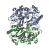
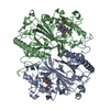
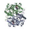
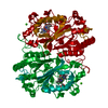
- PDB-6q3e: Dye type peroxidase Aa from Streptomyces lividans: 274.4 kGy structure -
+ Open data

- Basic information
| Entry | Database: PDB / ID: 6q3e | ||||||
|---|---|---|---|---|---|---|---|
| Title | Dye type peroxidase Aa from Streptomyces lividans: 274.4 kGy structure | ||||||
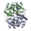
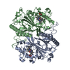
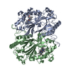
 Components Components | Deferrochelatase/peroxidase | ||||||
 Keywords Keywords |  OXIDOREDUCTASE / Dye type peroxidase / dose series / serial crystallography / heme OXIDOREDUCTASE / Dye type peroxidase / dose series / serial crystallography / heme | ||||||
| Function / homology |  Function and homology information Function and homology informationiron import into cell / ferrochelatase activity / Oxidoreductases; Acting on a peroxide as acceptor; Peroxidases / peroxidase activity / heme binding / metal ion binding / cytosolSimilarity search - Function | ||||||
| Biological species | Streptomyces coelicolor A3 | ||||||
| Method | X-RAY DIFFRACTION / SYNCHROTRON / MOLECULAR REPLACEMENT / Resolution: 2.03 Å | ||||||
 Authors Authors | Ebrahim, A. / Moreno-Chicano, T. / Worrall, J.A.R. / Strange, R.W. / Axford, D. / Sherrell, D.A. / Appleby, M. / Owen, R.L. | ||||||
 Citation Citation | Journal: Iucrj / Year: 2019 Title: Dose-resolved serial synchrotron and XFEL structures of radiation-sensitive metalloproteins. Authors: Ebrahim, A. / Moreno-Chicano, T. / Appleby, M.V. / Chaplin, A.K. / Beale, J.H. / Sherrell, D.A. / Duyvesteyn, H.M.E. / Owada, S. / Tono, K. / Sugimoto, H. / Strange, R.W. / Worrall, J.A.R. / ...Authors: Ebrahim, A. / Moreno-Chicano, T. / Appleby, M.V. / Chaplin, A.K. / Beale, J.H. / Sherrell, D.A. / Duyvesteyn, H.M.E. / Owada, S. / Tono, K. / Sugimoto, H. / Strange, R.W. / Worrall, J.A.R. / Axford, D. / Owen, R.L. / Hough, M.A. | ||||||
| History |
|
- Structure visualization
| Structure viewer | Molecule: MolmilJmol/JSmol |
|---|
- Downloads & links
-Download
| PDBx/mmCIF format | 6q3e.cif.gz | 158.3 KB | Display | PDBx/mmCIF format |
|---|---|---|---|---|
| PDB format | pdb6q3e.ent.gz | 123.1 KB | Display | PDB format |
| PDBx/mmJSON format | 6q3e.json.gz | Tree view | PDBx/mmJSON format | |
| Others |  Other downloads Other downloads |
-Validation report
| Arichive directory | https://data.pdbj.org/pub/pdb/validation_reports/q3/6q3eftp://data.pdbj.org/pub/pdb/validation_reports/q3/6q3e | HTTPS FTP |
|---|
-Related structure data
| Related structure data | 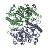 6i43SC 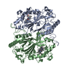 6i7zC  6i8eC  6i8iC  6i8jC  6i8kC  6i8oC  6i8pC 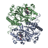 6i8qC  6ibnC  6q31C 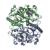 6q34C 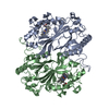 6q3dC S: Starting model for refinement C: citing same article ( |
|---|---|
| Similar structure data |


-Links
 PDBj
PDBj

- Assembly
| Deposited unit | 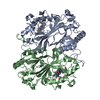
| ||||||||
|---|---|---|---|---|---|---|---|---|---|
| 1 |
| ||||||||
| Unit cell |
|
-Components
| #1: Protein | Mass: 39272.016 Da / Num. of mol.: 2 Source method: isolated from a genetically manipulated source Source: (gene. exp.)  Streptomyces coelicolor A3(2) (bacteria) Streptomyces coelicolor A3(2) (bacteria)Gene: SCO2276 / Production host: Escherichia coli (E. coli)References: UniProt: Q9RKQ2, Oxidoreductases; Acting on a peroxide as acceptor; Peroxidases#2: Chemical | Heme B  Mass: 616.487 Da / Num. of mol.: 2 Mass: 616.487 Da / Num. of mol.: 2Source method: isolated from a genetically manipulated source Formula: C34H32FeN4O4 #3: Water | ChemComp-HOH / | Water Mass: 18.015 Da / Num. of mol.: 211 / Source method: isolated from a natural source / Formula: H2O Mass: 18.015 Da / Num. of mol.: 211 / Source method: isolated from a natural source / Formula: H2O |
|---|
-Experimental details
-Experiment
| Experiment | Method: X-RAY DIFFRACTION / Number of used crystals: 1 |
|---|
- Sample preparation
| Crystal | Density Matthews: 2.28 Å3/Da / Density % sol: 46.16 % / Description: Microcrystals |
|---|---|
| Crystal grow | Temperature: 293 K / Method: batch mode / pH: 8 Details: 7 mg/ml protein mixed with 17% PEG 1500 and 67 mM MIB buffer (comprising imidazole, malonate and boric acid) |
-Data collection
| Diffraction | Mean temperature: 293 K / Serial crystal experiment: Y |
|---|---|
| Diffraction source | Source: SYNCHROTRON / Site: Diamond  / Beamline: I24 / Wavelength: 0.9686 Å / Beamline: I24 / Wavelength: 0.9686 Å |
| Detector | Type: DECTRIS PILATUS3 6M / Detector: PIXEL / Date: Jun 28, 2018 |
| Radiation | Monochromator: Silicon / Protocol: SINGLE WAVELENGTH / Monochromatic (M) / Laue (L): M / Scattering type: x-ray |
| Radiation wavelength | Wavelength: 0.9686 Å / Relative weight: 1 |
| Reflection | Resolution: 2.03→44.7 Å / Num. obs: 46140 / % possible obs: 100 % / Redundancy: 44.5 % / CC1/2: 0.969 / R split: 0.179 / Rmerge(I) obs: 0.765 / Net I/σ(I): 2.02 |
| Reflection shell | Resolution: 2.03→2.07 Å / Redundancy: 11.4 % / Rmerge(I) obs: 0.859 / Mean I/σ(I) obs: 0.5 / CC1/2: 0.506 / R split: 0.783 / % possible all: 99.6 |
| Serial crystallography measurement | Collection time total: 0.25 hours / Collimation: 2x Kirkpatrick-Baez mirror pairs |
| Serial crystallography sample delivery | Description: Silicon nitride chips / Method: fixed target |
- Processing
| Software |
| ||||||||||||||||||||||||||||||||||||||||||||||||||||||||||||||||||||||||||||||||||||||||||
|---|---|---|---|---|---|---|---|---|---|---|---|---|---|---|---|---|---|---|---|---|---|---|---|---|---|---|---|---|---|---|---|---|---|---|---|---|---|---|---|---|---|---|---|---|---|---|---|---|---|---|---|---|---|---|---|---|---|---|---|---|---|---|---|---|---|---|---|---|---|---|---|---|---|---|---|---|---|---|---|---|---|---|---|---|---|---|---|---|---|---|---|
| Refinement | Method to determine structure: MOLECULAR REPLACEMENT Starting model: 6I43 Resolution: 2.03→37.386 Å / SU ML: 0.32 / Cross valid method: THROUGHOUT / σ(F): 1.35 / Phase error: 24.83
| ||||||||||||||||||||||||||||||||||||||||||||||||||||||||||||||||||||||||||||||||||||||||||
| Solvent computation | Shrinkage radii: 0.9 Å / VDW probe radii: 1.11 Å | ||||||||||||||||||||||||||||||||||||||||||||||||||||||||||||||||||||||||||||||||||||||||||
| Displacement parameters | Biso max: 89.76 Å2 / Biso mean: 32.5936 Å2 / Biso min: 11.7 Å2 | ||||||||||||||||||||||||||||||||||||||||||||||||||||||||||||||||||||||||||||||||||||||||||
| Refinement step | Cycle: final / Resolution: 2.03→37.386 Å
| ||||||||||||||||||||||||||||||||||||||||||||||||||||||||||||||||||||||||||||||||||||||||||
| LS refinement shell | Refine-ID: X-RAY DIFFRACTION / Rfactor Rfree error: 0 / Total num. of bins used: 14 / % reflection obs: 100 %
|
