 Movie
Movie Controller

[English] 日本語
 Yorodumi
- PDB-5yzc: Crystal structure of the prefusion form of measles virus fusion p... -
+ Open data

- Basic information
| Entry | Database: PDB / ID: 5yzc | ||||||||||||||||||
|---|---|---|---|---|---|---|---|---|---|---|---|---|---|---|---|---|---|---|---|
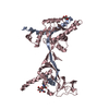
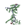
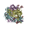
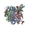
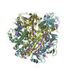
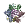
| Title | Crystal structure of the prefusion form of measles virus fusion protein in complex with a fusion inhibitor compound (AS-48) | ||||||||||||||||||
 Components Components | (glycoprotein ... ) x 2 ) x 2 | ||||||||||||||||||
 Keywords Keywords | VIRAL PROTEIN / glycoprotein / fusion protein / paramyxovirus / inhibitor / chemical compound | ||||||||||||||||||
| Function / homology |  Function and homology information Function and homology informationsymbiont entry into host cell / fusion of virus membrane with host plasma membrane / viral envelope / host cell plasma membrane / virion membrane / membraneSimilarity search - Function | ||||||||||||||||||
| Biological species |   Measles virus Measles virus | ||||||||||||||||||
| Method | X-RAY DIFFRACTION / SYNCHROTRON / MOLECULAR REPLACEMENT / Resolution: 2.334 Å | ||||||||||||||||||
 Authors Authors | Hashiguchi, T. / Fukuda, Y. / Matsuoka, R. / Kuroda, D. / Kubota, M. / Shirogane, Y. / Watanabe, S. / Tsumoto, K. / Kohda, D. / Plemper, R.K. / Yanagi, Y. | ||||||||||||||||||
| Funding support |  Japan, Japan,  United States, 5items United States, 5items
| ||||||||||||||||||
 Citation Citation | Journal: Proc. Natl. Acad. Sci. U.S.A. / Year: 2018 Title: Structures of the prefusion form of measles virus fusion protein in complex with inhibitors. Authors: Hashiguchi, T. / Fukuda, Y. / Matsuoka, R. / Kuroda, D. / Kubota, M. / Shirogane, Y. / Watanabe, S. / Tsumoto, K. / Kohda, D. / Plemper, R.K. / Yanagi, Y. | ||||||||||||||||||
| History |
|
- Structure visualization
| Structure viewer | Molecule: MolmilJmol/JSmol |
|---|
- Downloads & links
-Download
| PDBx/mmCIF format | 5yzc.cif.gz | 108.1 KB | Display | PDBx/mmCIF format |
|---|---|---|---|---|
| PDB format | pdb5yzc.ent.gz | 79 KB | Display | PDB format |
| PDBx/mmJSON format | 5yzc.json.gz | Tree view | PDBx/mmJSON format | |
| Others |  Other downloads Other downloads |
-Validation report
| Arichive directory | https://data.pdbj.org/pub/pdb/validation_reports/yz/5yzcftp://data.pdbj.org/pub/pdb/validation_reports/yz/5yzc | HTTPS FTP |
|---|
-Related structure data
| Related structure data |  5yxwSC  5yzdC S: Starting model for refinement C: citing same article ( |
|---|---|
| Similar structure data |


-Links
 PDBj
PDBj

- Assembly
| Deposited unit | 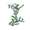
| ||||||||
|---|---|---|---|---|---|---|---|---|---|
| 1 | 
| ||||||||
| Unit cell |
|
-Components
-Glycoprotein ... , 2 types, 2 molecules AB
| #1: Protein | Mass: 10524.273 Da / Num. of mol.: 1 Source method: isolated from a genetically manipulated source Source: (gene. exp.) Measles virus (strain Ichinose-B95a) / Strain: Ichinose-B95a / Gene: F / Cell (production host): S2 / Production host:  Drosophila melanogaster (fruit fly) / References: UniProt: Q786F3 Drosophila melanogaster (fruit fly) / References: UniProt: Q786F3 |
|---|---|
| #2: Protein | Mass: 44925.941 Da / Num. of mol.: 1 Source method: isolated from a genetically manipulated source Details: The fusion protein of glycoprotein F1,measles virus fusion protein and Tags Source: (gene. exp.) Measles virus (strain Ichinose-B95a), (gene. exp.) Measles virusStrain: Ichinose-B95a, IC-B / Gene: F / Cell (production host): S2 / Production host: Drosophila melanogaster (fruit fly) / References: UniProt: Q786F3 |
-Sugars , 2 types, 2 molecules 
| #3: Polysaccharide | 2-acetamido-2-deoxy-beta-D-glucopyranose-(1-4)-2-acetamido-2-deoxy-beta-D-glucopyranose / Mass: 424.401 Da / Num. of mol.: 1 Source method: isolated from a genetically manipulated source |
|---|---|
| #4: Sugar | ChemComp-NAG / N-Acetylglucosamine Type: D-saccharide, beta linking / Mass: 221.208 Da / Num. of mol.: 1 Type: D-saccharide, beta linking / Mass: 221.208 Da / Num. of mol.: 1Source method: isolated from a genetically manipulated source Formula: C8H15NO6 |
-Non-polymers , 2 types, 159 molecules 

| #5: Chemical | ChemComp-95C /  Mass: 299.281 Da / Num. of mol.: 1 / Source method: obtained synthetically / Formula: C15H13N3O4 Mass: 299.281 Da / Num. of mol.: 1 / Source method: obtained synthetically / Formula: C15H13N3O4 |
|---|---|
| #6: Water | ChemComp-HOH / WaterMass: 18.015 Da / Num. of mol.: 158 / Source method: isolated from a natural source / Formula: H2O |
-Experimental details
-Experiment
| Experiment | Method: X-RAY DIFFRACTION / Number of used crystals: 1 |
|---|
- Sample preparation
| Crystal | Density Matthews: 3.66 Å3/Da / Density % sol: 66.38 % |
|---|---|
| Crystal grow | Temperature: 293 K / Method: vapor diffusion, hanging drop / Details: sodium formate, glycerol, sodium acetate |
-Data collection
| Diffraction | Mean temperature: 100 K |
|---|---|
| Diffraction source | Source: SYNCHROTRON / Site: Photon Factory / Beamline: BL-1A / Wavelength: 1.1 Å |
| Detector | Type: DECTRIS EIGER X 4M / Detector: PIXEL / Date: Feb 23, 2017 |
| Radiation | Protocol: SINGLE WAVELENGTH / Monochromatic (M) / Laue (L): M / Scattering type: x-ray |
| Radiation wavelength | Wavelength: 1.1 Å / Relative weight: 1 |
| Reflection | Resolution: 2.334→119.852 Å / Num. obs: 34515 / % possible obs: 100 % / Redundancy: 20.9 % / Net I/σ(I): 14.3 |
| Reflection shell | Resolution: 2.334→2.342 Å |
- Processing
| Software |
| |||||||||||||||||||||||||||||||||||||||||||||||||||||||||||||||||||||||||||||||||||||||||||
|---|---|---|---|---|---|---|---|---|---|---|---|---|---|---|---|---|---|---|---|---|---|---|---|---|---|---|---|---|---|---|---|---|---|---|---|---|---|---|---|---|---|---|---|---|---|---|---|---|---|---|---|---|---|---|---|---|---|---|---|---|---|---|---|---|---|---|---|---|---|---|---|---|---|---|---|---|---|---|---|---|---|---|---|---|---|---|---|---|---|---|---|---|
| Refinement | Method to determine structure: MOLECULAR REPLACEMENT Starting model: 5YXW Resolution: 2.334→53.6 Å / SU ML: 0.22 / Cross valid method: FREE R-VALUE / σ(F): 1.34 / Phase error: 21.76 / Stereochemistry target values: ML
| |||||||||||||||||||||||||||||||||||||||||||||||||||||||||||||||||||||||||||||||||||||||||||
| Solvent computation | Shrinkage radii: 0.9 Å / VDW probe radii: 1.11 Å / Solvent model: FLAT BULK SOLVENT MODEL | |||||||||||||||||||||||||||||||||||||||||||||||||||||||||||||||||||||||||||||||||||||||||||
| Refinement step | Cycle: LAST / Resolution: 2.334→53.6 Å
| |||||||||||||||||||||||||||||||||||||||||||||||||||||||||||||||||||||||||||||||||||||||||||
| Refine LS restraints |
| |||||||||||||||||||||||||||||||||||||||||||||||||||||||||||||||||||||||||||||||||||||||||||
| LS refinement shell |
|
