 Movie
Movie Controller

[English] 日本語
 Yorodumi
- PDB-5evm: Crystal Structure of Nipah Virus Fusion Glycoprotein in the Prefu... -
+ Open data

- Basic information
| Entry | Database: PDB / ID: 5evm | ||||||||||||
|---|---|---|---|---|---|---|---|---|---|---|---|---|---|
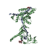
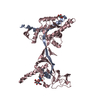
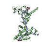
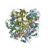
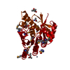
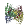
| Title | Crystal Structure of Nipah Virus Fusion Glycoprotein in the Prefusion State | ||||||||||||
 Components Components | Fusion glycoprotein F0 | ||||||||||||
 Keywords Keywords |  VIRAL PROTEIN / fusion protein / Nipah / prefusion / paramyxovirus / henipavirus VIRAL PROTEIN / fusion protein / Nipah / prefusion / paramyxovirus / henipavirus | ||||||||||||
| Function / homology |  Function and homology information Function and homology informationmembrane fusion involved in viral entry into host cell / symbiont entry into host cell / fusion of virus membrane with host plasma membrane / viral envelope / host cell plasma membrane / virion membrane / membraneSimilarity search - Function | ||||||||||||
| Biological species |  Nipah virus Nipah virus | ||||||||||||
| Method | X-RAY DIFFRACTION / SYNCHROTRON / MOLECULAR REPLACEMENT / Resolution: 3.367 Å | ||||||||||||
 Authors Authors | Xu, K. / Nikolov, D.B. | ||||||||||||
| Funding support |  United States, 3items United States, 3items
| ||||||||||||
 Citation Citation | Journal: Plos Pathog. / Year: 2015 Title: Crystal Structure of the Pre-fusion Nipah Virus Fusion Glycoprotein Reveals a Novel Hexamer-of-Trimers Assembly. Authors: Xu, K. / Chan, Y.P. / Bradel-Tretheway, B. / Akyol-Ataman, Z. / Zhu, Y. / Dutta, S. / Yan, L. / Feng, Y. / Wang, L.F. / Skiniotis, G. / Lee, B. / Zhou, Z.H. / Broder, C.C. / Aguilar, H.C. / Nikolov, D.B. | ||||||||||||
| History |
|
- Structure visualization
| Structure viewer | Molecule: MolmilJmol/JSmol |
|---|
- Downloads & links
-Download
| PDBx/mmCIF format | 5evm.cif.gz | 494.5 KB | Display | PDBx/mmCIF format |
|---|---|---|---|---|
| PDB format | pdb5evm.ent.gz | 413.9 KB | Display | PDB format |
| PDBx/mmJSON format | 5evm.json.gz | Tree view | PDBx/mmJSON format | |
| Others |  Other downloads Other downloads |
-Validation report
| Arichive directory | https://data.pdbj.org/pub/pdb/validation_reports/ev/5evmftp://data.pdbj.org/pub/pdb/validation_reports/ev/5evm | HTTPS FTP |
|---|
-Related structure data
| Related structure data | 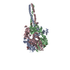 2b9bS S: Starting model for refinement |
|---|---|
| Similar structure data |


-Links
 PDBj
PDBj

- Assembly
| Deposited unit | 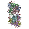
| ||||||||
|---|---|---|---|---|---|---|---|---|---|
| 1 | 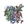
| ||||||||
| 2 | 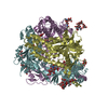
| ||||||||
| Unit cell |
|
-Components
-Protein / Non-polymers , 2 types, 12 molecules ABCDEF
| #1: Protein | Mass: 58407.285 Da / Num. of mol.: 6 / Mutation: N67D, N305D Source method: isolated from a genetically manipulated source Source: (gene. exp.) Nipah virus / Cell (production host): Epithelia / Organ (production host): KIDNEY / Production host:  Homo sapiens (human) / References: UniProt: Q9IH63 Homo sapiens (human) / References: UniProt: Q9IH63#9: Chemical | ChemComp-MLI / Malonic acid Mass: 102.046 Da / Num. of mol.: 6 / Source method: obtained synthetically / Formula: C3H2O4 Mass: 102.046 Da / Num. of mol.: 6 / Source method: obtained synthetically / Formula: C3H2O4 |
|---|
-Sugars , 7 types, 18 molecules
| #2: Polysaccharide | alpha-D-mannopyranose-(1-2)-[alpha-D-mannopyranose-(1-6)]beta-D-mannopyranose-(1-4)-2-acetamido-2- ...alpha-D-mannopyranose-(1-2)-[alpha-D-mannopyranose-(1-6)]beta-D-mannopyranose-(1-4)-2-acetamido-2-deoxy-beta-D-glucopyranose-(1-4)-2-acetamido-2-deoxy-beta-D-glucopyranose / Mass: 910.823 Da / Num. of mol.: 1 Source method: isolated from a genetically manipulated source | ||||||||||
|---|---|---|---|---|---|---|---|---|---|---|---|
| #3: Polysaccharide | 2-acetamido-2-deoxy-beta-D-glucopyranose-(1-4)-2-acetamido-2-deoxy-beta-D-glucopyranose / Mass: 424.401 Da / Num. of mol.: 5Source method: isolated from a genetically manipulated source #4: Polysaccharide | alpha-D-mannopyranose-(1-4)-2-acetamido-2-deoxy-beta-D-glucopyranose-(1-4)-2-acetamido-2-deoxy-beta- ...alpha-D-mannopyranose-(1-4)-2-acetamido-2-deoxy-beta-D-glucopyranose-(1-4)-2-acetamido-2-deoxy-beta-D-glucopyranose / Mass: 586.542 Da / Num. of mol.: 6Source method: isolated from a genetically manipulated source #5: Polysaccharide | / Mass: 910.823 Da / Num. of mol.: 2Source method: isolated from a genetically manipulated source #6: Polysaccharide | alpha-D-mannopyranose-(1-2)-[alpha-D-mannopyranose-(1-6)]alpha-D-mannopyranose-(1-4)-2-acetamido-2- ...alpha-D-mannopyranose-(1-2)-[alpha-D-mannopyranose-(1-6)]alpha-D-mannopyranose-(1-4)-2-acetamido-2-deoxy-beta-D-glucopyranose-(1-4)-2-acetamido-2-deoxy-beta-D-glucopyranose | / Mass: 910.823 Da / Num. of mol.: 1Source method: isolated from a genetically manipulated source #7: Polysaccharide | alpha-D-mannopyranose-(1-6)-alpha-D-mannopyranose-(1-4)-2-acetamido-2-deoxy-beta-D-glucopyranose-(1- ...alpha-D-mannopyranose-(1-6)-alpha-D-mannopyranose-(1-4)-2-acetamido-2-deoxy-beta-D-glucopyranose-(1-4)-2-acetamido-2-deoxy-beta-D-glucopyranose | / Mass: 748.682 Da / Num. of mol.: 1Source method: isolated from a genetically manipulated source #8: Polysaccharide | / Mass: 748.682 Da / Num. of mol.: 2Source method: isolated from a genetically manipulated source |
-Experimental details
-Experiment
| Experiment | Method: X-RAY DIFFRACTION |
|---|
- Sample preparation
| Crystal | Density Matthews: 5.87 Å3/Da / Density % sol: 79.04 % |
|---|---|
| Crystal grow | Temperature: 298 K / Method: vapor diffusion, hanging drop / pH: 7.3 Details: 0.1 M HEPES, 1% Jeffamine ED-2001, 1.2 M sodium malonate |
-Data collection
| Diffraction | Mean temperature: 100 K |
|---|---|
| Diffraction source | Source: SYNCHROTRON / Site: APS / Beamline: 24-ID-C / Wavelength: 0.9792 Å |
| Detector | Type: DECTRIS PILATUS 6M / Detector: PIXEL / Date: Mar 1, 2011 |
| Radiation | Monochromator: double crystal Si(111) / Protocol: SINGLE WAVELENGTH / Monochromatic (M) / Laue (L): M / Scattering type: x-ray |
| Radiation wavelength | Wavelength: 0.9792 Å / Relative weight: 1 |
| Reflection | Resolution: 3.35→50 Å / Num. all: 112890 / Num. obs: 112120 / % possible obs: 99.36 % / Redundancy: 5.9 % / Rmerge(I) obs: 0.161 / Net I/σ(I): 2.41 |
| Reflection shell | Resolution: 3.35→3.47 Å / Redundancy: 5.5 % / Rmerge(I) obs: 0.975 / Mean I/σ(I) obs: 2.41 / % possible all: 93.93 |
- Processing
| Software |
| |||||||||||||||||||||||||||||||||||||||||||||||||||||||||||||||||||||||||||||||||||||||||||||||||||||||||||||||||||||||||||||||||||||||||||||||||||||||||||||||||||||||||||||||||||||||||||||||||||||||||||||||||||||||||
|---|---|---|---|---|---|---|---|---|---|---|---|---|---|---|---|---|---|---|---|---|---|---|---|---|---|---|---|---|---|---|---|---|---|---|---|---|---|---|---|---|---|---|---|---|---|---|---|---|---|---|---|---|---|---|---|---|---|---|---|---|---|---|---|---|---|---|---|---|---|---|---|---|---|---|---|---|---|---|---|---|---|---|---|---|---|---|---|---|---|---|---|---|---|---|---|---|---|---|---|---|---|---|---|---|---|---|---|---|---|---|---|---|---|---|---|---|---|---|---|---|---|---|---|---|---|---|---|---|---|---|---|---|---|---|---|---|---|---|---|---|---|---|---|---|---|---|---|---|---|---|---|---|---|---|---|---|---|---|---|---|---|---|---|---|---|---|---|---|---|---|---|---|---|---|---|---|---|---|---|---|---|---|---|---|---|---|---|---|---|---|---|---|---|---|---|---|---|---|---|---|---|---|---|---|---|---|---|---|---|---|---|---|---|---|---|---|---|---|
| Refinement | Method to determine structure: MOLECULAR REPLACEMENT Starting model: PDB entry 2B9B Resolution: 3.367→43.158 Å / SU ML: 0.43 / Cross valid method: FREE R-VALUE / σ(F): 1.96 / Phase error: 28.24 / Stereochemistry target values: ML
| |||||||||||||||||||||||||||||||||||||||||||||||||||||||||||||||||||||||||||||||||||||||||||||||||||||||||||||||||||||||||||||||||||||||||||||||||||||||||||||||||||||||||||||||||||||||||||||||||||||||||||||||||||||||||
| Solvent computation | Shrinkage radii: 0.9 Å / VDW probe radii: 1.11 Å / Solvent model: FLAT BULK SOLVENT MODEL | |||||||||||||||||||||||||||||||||||||||||||||||||||||||||||||||||||||||||||||||||||||||||||||||||||||||||||||||||||||||||||||||||||||||||||||||||||||||||||||||||||||||||||||||||||||||||||||||||||||||||||||||||||||||||
| Refinement step | Cycle: LAST / Resolution: 3.367→43.158 Å
| |||||||||||||||||||||||||||||||||||||||||||||||||||||||||||||||||||||||||||||||||||||||||||||||||||||||||||||||||||||||||||||||||||||||||||||||||||||||||||||||||||||||||||||||||||||||||||||||||||||||||||||||||||||||||
| Refine LS restraints |
| |||||||||||||||||||||||||||||||||||||||||||||||||||||||||||||||||||||||||||||||||||||||||||||||||||||||||||||||||||||||||||||||||||||||||||||||||||||||||||||||||||||||||||||||||||||||||||||||||||||||||||||||||||||||||
| LS refinement shell |
|
