 Movie
Movie Controller

[English] 日本語
 Yorodumi
- PDB-5ydi: Crystal structure of acetylcholinesterase catalytic subunits of t... -
+ Open data

- Basic information
| Entry | Database: PDB / ID: 5ydi | |||||||||
|---|---|---|---|---|---|---|---|---|---|---|
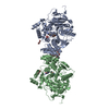
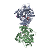
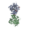
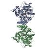
| Title | Crystal structure of acetylcholinesterase catalytic subunits of the malaria vector anopheles gambiae, new crystal packing | |||||||||
 Components Components | Acetylcholinesterase | |||||||||
 Keywords Keywords | HYDROLASE / ALPHA/BATA HYDROLASE | |||||||||
| Function / homology |  Function and homology information Function and homology informationacetylcholine catabolic process / acetylcholinesterase / acetylcholinesterase activity / choline metabolic process / synapse / extracellular space / plasma membraneSimilarity search - Function | |||||||||
| Biological species |  Anopheles gambiae (African malaria mosquito) Anopheles gambiae (African malaria mosquito) | |||||||||
| Method | X-RAY DIFFRACTION / SYNCHROTRON / MOLECULAR REPLACEMENT / Resolution: 3.45 Å | |||||||||
 Authors Authors | Han, Q. / Guan, H. / Ding, H. / Liao, C. / Robinson, H. / Li, J. | |||||||||
| Funding support |  China, China,  United States, 2items United States, 2items
| |||||||||
 Citation Citation | Journal: To Be Published Title: Crystal structures of acetylcholinesterase of the malaria vector Anopheles gambiae reveal a polymerization interface, ligand binding residues and post translational modifications Authors: Han, Q. / Guan, H. / Ding, H. / Liao, C. / Robinson, H. / Li, J. | |||||||||
| History |
|
- Structure visualization
| Structure viewer | Molecule: MolmilJmol/JSmol |
|---|
- Downloads & links
-Download
| PDBx/mmCIF format | 5ydi.cif.gz | 325 KB | Display | PDBx/mmCIF format |
|---|---|---|---|---|
| PDB format | pdb5ydi.ent.gz | 263.7 KB | Display | PDB format |
| PDBx/mmJSON format | 5ydi.json.gz | Tree view | PDBx/mmJSON format | |
| Others |  Other downloads Other downloads |
-Validation report
| Arichive directory | https://data.pdbj.org/pub/pdb/validation_reports/yd/5ydiftp://data.pdbj.org/pub/pdb/validation_reports/yd/5ydi | HTTPS FTP |
|---|
-Related structure data
| Related structure data |  5ydhSC  5ydjC S: Starting model for refinement C: citing same article ( |
|---|---|
| Similar structure data |


-Links
 PDBj
PDBj
- Assembly
| Deposited unit | 
| ||||||||
|---|---|---|---|---|---|---|---|---|---|
| 1 | 
| ||||||||
| 2 | 
| ||||||||
| Unit cell |
|
-Components
-Protein , 1 types, 3 molecules ABC
| #1: Protein | / AChE Mass: 61684.125 Da / Num. of mol.: 3 / Fragment: CATALYTIC SUBUNIT, UNP RESIDUES 162-714 Source method: isolated from a genetically manipulated source Source: (gene. exp.) Anopheles gambiae (African malaria mosquito)Gene: Ace, ACE1, ACHE1, AGAP001356 / Plasmid: PPICZ*A / Production host:  Komagataella pastoris (fungus) / References: UniProt: Q869C3, acetylcholinesterase Komagataella pastoris (fungus) / References: UniProt: Q869C3, acetylcholinesterase |
|---|
-Sugars , 3 types, 6 molecules 
| #2: Polysaccharide | / Mass: 586.542 Da / Num. of mol.: 2 Source method: isolated from a genetically manipulated source #3: Polysaccharide | alpha-D-mannopyranose-(1-6)-alpha-D-mannopyranose-(1-3)-[alpha-D-mannopyranose-(1-6)]beta-D- ...alpha-D-mannopyranose-(1-6)-alpha-D-mannopyranose-(1-3)-[alpha-D-mannopyranose-(1-6)]beta-D-mannopyranose-(1-4)-2-acetamido-2-deoxy-beta-D-glucopyranose-(1-4)-2-acetamido-2-deoxy-beta-D-glucopyranose | / Mass: 1072.964 Da / Num. of mol.: 1Source method: isolated from a genetically manipulated source #4: Sugar | N-Acetylglucosamine Type: D-saccharide, beta linking / Mass: 221.208 Da / Num. of mol.: 3 Type: D-saccharide, beta linking / Mass: 221.208 Da / Num. of mol.: 3Source method: isolated from a genetically manipulated source Formula: C8H15NO6 |
|---|
-Non-polymers , 3 types, 18 molecules 


| #5: Chemical | ChemComp-GOL / Glycerol Mass: 92.094 Da / Num. of mol.: 4 / Source method: obtained synthetically / Formula: C3H8O3 Mass: 92.094 Da / Num. of mol.: 4 / Source method: obtained synthetically / Formula: C3H8O3#6: Chemical | ChemComp-NA /  Mass: 22.990 Da / Num. of mol.: 4 / Source method: obtained synthetically / Formula: Na Mass: 22.990 Da / Num. of mol.: 4 / Source method: obtained synthetically / Formula: Na#7: Water | ChemComp-HOH / | WaterMass: 18.015 Da / Num. of mol.: 10 / Source method: isolated from a natural source / Formula: H2O |
|---|
-Experimental details
-Experiment
| Experiment | Method: X-RAY DIFFRACTION / Number of used crystals: 1 |
|---|
- Sample preparation
| Crystal | Density Matthews: 3.4 Å3/Da / Density % sol: 63.79 % |
|---|---|
| Crystal grow | Temperature: 277 K / Method: vapor diffusion, hanging drop / pH: 7.5 Details: 0.1M HEPES BUFFER, 20% PEG 4000, 22% GLYCEROL, PH 7.5, VAPOR DIFFUSION, HANGING DROP, TEMPERATURE 277K |
-Data collection
| Diffraction | Mean temperature: 100 K |
|---|---|
| Diffraction source | Source: SYNCHROTRON / Site: NSLS / Beamline: X29A / Wavelength: 1.075 Å |
| Detector | Type: ADSC QUANTUM 315 / Detector: CCD / Date: May 28, 2013 |
| Radiation | Monochromator: SI 111 CHANNEL / Protocol: SINGLE WAVELENGTH / Monochromatic (M) / Laue (L): M / Scattering type: x-ray |
| Radiation wavelength | Wavelength: 1.075 Å / Relative weight: 1 |
| Reflection | Resolution: 3.45→63.72 Å / Num. obs: 31768 / % possible obs: 76 % / Observed criterion σ(I): -3 / Redundancy: 13.1 % / Net I/σ(I): 11.7 |
| Reflection shell | Resolution: 3.45→3.64 Å |
- Processing
| Software |
| ||||||||||||||||||||||||||||||||||||||||||||||||||||||||||||||||||||||||||||||||||||||||||||||||||||||||||||||||||||||||||||||||||||||||||||||||||||||||||||||||||||||||||||||||||||||
|---|---|---|---|---|---|---|---|---|---|---|---|---|---|---|---|---|---|---|---|---|---|---|---|---|---|---|---|---|---|---|---|---|---|---|---|---|---|---|---|---|---|---|---|---|---|---|---|---|---|---|---|---|---|---|---|---|---|---|---|---|---|---|---|---|---|---|---|---|---|---|---|---|---|---|---|---|---|---|---|---|---|---|---|---|---|---|---|---|---|---|---|---|---|---|---|---|---|---|---|---|---|---|---|---|---|---|---|---|---|---|---|---|---|---|---|---|---|---|---|---|---|---|---|---|---|---|---|---|---|---|---|---|---|---|---|---|---|---|---|---|---|---|---|---|---|---|---|---|---|---|---|---|---|---|---|---|---|---|---|---|---|---|---|---|---|---|---|---|---|---|---|---|---|---|---|---|---|---|---|---|---|---|---|
| Refinement | Method to determine structure: MOLECULAR REPLACEMENT Starting model: 5YDH Resolution: 3.45→63.72 Å / Cor.coef. Fo:Fc: 0.906 / Cor.coef. Fo:Fc free: 0.854 / SU B: 25.984 / SU ML: 0.404 / Cross valid method: THROUGHOUT / ESU R Free: 0.543
| ||||||||||||||||||||||||||||||||||||||||||||||||||||||||||||||||||||||||||||||||||||||||||||||||||||||||||||||||||||||||||||||||||||||||||||||||||||||||||||||||||||||||||||||||||||||
| Solvent computation | Ion probe radii: 0.8 Å / Shrinkage radii: 0.8 Å / VDW probe radii: 1.2 Å | ||||||||||||||||||||||||||||||||||||||||||||||||||||||||||||||||||||||||||||||||||||||||||||||||||||||||||||||||||||||||||||||||||||||||||||||||||||||||||||||||||||||||||||||||||||||
| Displacement parameters | Biso mean: 62.64 Å2
| ||||||||||||||||||||||||||||||||||||||||||||||||||||||||||||||||||||||||||||||||||||||||||||||||||||||||||||||||||||||||||||||||||||||||||||||||||||||||||||||||||||||||||||||||||||||
| Refinement step | Cycle: LAST / Resolution: 3.45→63.72 Å
| ||||||||||||||||||||||||||||||||||||||||||||||||||||||||||||||||||||||||||||||||||||||||||||||||||||||||||||||||||||||||||||||||||||||||||||||||||||||||||||||||||||||||||||||||||||||
| Refine LS restraints |
|