 Movie
Movie Controller

[English] 日本語
 Yorodumi
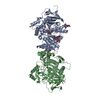
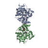
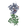
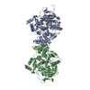
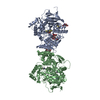
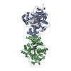
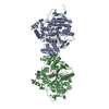
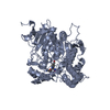
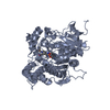
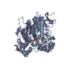
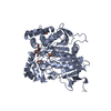
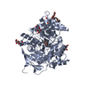
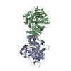
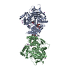
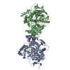
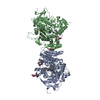
- PDB-2jgm: Crystal structure of mouse acetylcholinesterase inhibited by aged... -
+ Open data

- Basic information
| Entry | Database: PDB / ID: 2jgm | ||||||
|---|---|---|---|---|---|---|---|
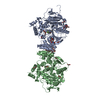
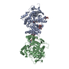
| Title | Crystal structure of mouse acetylcholinesterase inhibited by aged diisopropyl fluorophosphate (DFP) | ||||||
 Components Components | ACETYLCHOLINESTERASE | ||||||
 Keywords Keywords | HYDROLASE / NEUROTRANSMITTER DEGRADATION / DFP / AGING / SYNAPSE / MEMBRANE / GLYCOPROTEIN / SERINE ESTERASE / ACETYLCHOLINESTERASE / ALTERNATIVE SPLICING / DIISOPROPYL FLUOROPHOSPHATE | ||||||
| Function / homology |  Function and homology information Function and homology informationacetylcholine metabolic process / serine hydrolase activity / choline binding / acetylcholine catabolic process / acetylcholine binding / acetylcholinesterase / acetylcholine receptor signaling pathway / positive regulation of dendrite morphogenesis / osteoblast development / acetylcholinesterase activity ...acetylcholine metabolic process / serine hydrolase activity / choline binding / acetylcholine catabolic process / acetylcholine binding / acetylcholinesterase / acetylcholine receptor signaling pathway / positive regulation of dendrite morphogenesis / osteoblast development / acetylcholinesterase activity / choline metabolic process / positive regulation of axonogenesis / basement membrane / regulation of receptor recycling / laminin binding / side of membrane / synaptic cleft / synapse assembly / collagen binding / response to insulin / neuromuscular junction / receptor internalization / : / retina development in camera-type eye / nuclear envelope / presynaptic membrane / positive regulation of cold-induced thermogenesis / postsynaptic membrane / cell adhesion / endoplasmic reticulum lumen / axon / neuronal cell body / synapse / dendrite / perinuclear region of cytoplasm / Golgi apparatus / cell surface / protein homodimerization activity / extracellular space / identical protein binding / plasma membraneSimilarity search - Function | ||||||
| Biological species |  MUS MUSCULUS (house mouse) MUS MUSCULUS (house mouse) | ||||||
| Method | X-RAY DIFFRACTION / SYNCHROTRON / MOLECULAR REPLACEMENT / Resolution: 2.9 Å | ||||||
 Authors Authors | Hornberg, A. / Tunemalm, A.-K. / Ekstrom, F. | ||||||
 Citation Citation | Journal: Biochemistry / Year: 2007 Title: Crystal Structures of Acetylcholinesterase in Complex with Organophosphorus Compounds Suggest that the Acyl Pocket Modulates the Aging Reaction by Precluding the Formation of the Trigonal ...Title: Crystal Structures of Acetylcholinesterase in Complex with Organophosphorus Compounds Suggest that the Acyl Pocket Modulates the Aging Reaction by Precluding the Formation of the Trigonal Bipyramidal Transition State. Authors: Hornberg, A. / Tunemalm, A.-K. / Ekstrom, F. | ||||||
| History |
|
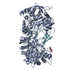
- Structure visualization
| Structure viewer | Molecule: MolmilJmol/JSmol |
|---|
- Downloads & links
-Download
| PDBx/mmCIF format | 2jgm.cif.gz | 215.3 KB | Display | PDBx/mmCIF format |
|---|---|---|---|---|
| PDB format | pdb2jgm.ent.gz | 173 KB | Display | PDB format |
| PDBx/mmJSON format | 2jgm.json.gz | Tree view | PDBx/mmJSON format | |
| Others |  Other downloads Other downloads |
-Validation report
| Arichive directory | https://data.pdbj.org/pub/pdb/validation_reports/jg/2jgmftp://data.pdbj.org/pub/pdb/validation_reports/jg/2jgm | HTTPS FTP |
|---|
-Related structure data
| Related structure data | 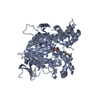 2jgeC 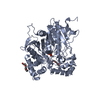 2jgfC 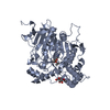 2jgiC 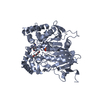 2jgjC 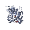 2jgkC 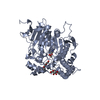 2jglC  1j06S S: Starting model for refinement C: citing same article ( |
|---|---|
| Similar structure data |


-Links
 PDBj
PDBj
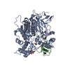
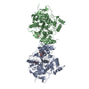
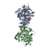
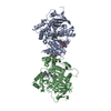
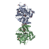
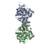
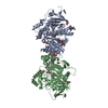
- Assembly
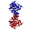
| Deposited unit | 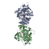
| ||||||||
|---|---|---|---|---|---|---|---|---|---|
| 1 | 
| ||||||||
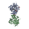
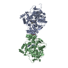
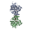
| Unit cell |
|
-Components
| #1: Protein | / ACHE Mass: 60356.047 Da / Num. of mol.: 2 / Fragment: CATALYTIC DOMAIN, RESIDUES 32-574 Source method: isolated from a genetically manipulated source Source: (gene. exp.) MUS MUSCULUS (house mouse) / Cell line (production host): HEK293F / Production host:  HOMO SAPIENS (human) / References: UniProt: P21836, acetylcholinesterase HOMO SAPIENS (human) / References: UniProt: P21836, acetylcholinesterase#2: Sugar | ChemComp-NAG / | N-Acetylglucosamine  Type: D-saccharide, beta linking / Mass: 221.208 Da / Num. of mol.: 1 Type: D-saccharide, beta linking / Mass: 221.208 Da / Num. of mol.: 1Source method: isolated from a genetically manipulated source Formula: C8H15NO6 #3: Water | ChemComp-HOH / | Water Mass: 18.015 Da / Num. of mol.: 92 / Source method: isolated from a natural source / Formula: H2O Mass: 18.015 Da / Num. of mol.: 92 / Source method: isolated from a natural source / Formula: H2OSequence details | GAP BETWEEN RESIDUES 257 AND 265 (MONOMER A AND B) MONOMER B STARTS AT RESIDUE 4 | |
|---|
-Experimental details
-Experiment
| Experiment | Method: X-RAY DIFFRACTION / Number of used crystals: 1 |
|---|
- Sample preparation
| Crystal | Density Matthews: 3.9 Å3/Da / Density % sol: 68 % |
|---|---|
| Crystal grow | pH: 7 / Details: 28% PEG 750MME, 0.1 M HEPES PH7.0, pH 7.00 |
-Data collection
| Diffraction | Mean temperature: 100 K |
|---|---|
| Diffraction source | Source: SYNCHROTRON / Site: MAX II  / Beamline: I911-5 / Wavelength: 0.906 / Beamline: I911-5 / Wavelength: 0.906 |
| Detector | Type: MARRESEARCH / Detector: CCD / Date: Apr 4, 2006 / Details: MIRRORS |
| Radiation | Monochromator: SI111 / Protocol: SINGLE WAVELENGTH / Monochromatic (M) / Laue (L): M / Scattering type: x-ray |
| Radiation wavelength | Wavelength: 0.906 Å / Relative weight: 1 |
| Reflection | Resolution: 2.9→29.89 Å / Num. obs: 45500 / % possible obs: 99.9 % / Observed criterion σ(I): 0 / Redundancy: 7.4 % / Rmerge(I) obs: 0.09 / Net I/σ(I): 19.8 |
| Reflection shell | Resolution: 2.9→3.06 Å / Redundancy: 7.5 % / Rmerge(I) obs: 0.37 / Mean I/σ(I) obs: 5.6 / % possible all: 100 |
- Processing
| Software |
| ||||||||||||||||||||||||||||||||||||||||||||||||||||||||||||||||||||||||||||||||||||||||||||||||||||||||||||||||||||||||||||||||||||||||||||||||||||||||||||||||||||||||||||||||||||||
|---|---|---|---|---|---|---|---|---|---|---|---|---|---|---|---|---|---|---|---|---|---|---|---|---|---|---|---|---|---|---|---|---|---|---|---|---|---|---|---|---|---|---|---|---|---|---|---|---|---|---|---|---|---|---|---|---|---|---|---|---|---|---|---|---|---|---|---|---|---|---|---|---|---|---|---|---|---|---|---|---|---|---|---|---|---|---|---|---|---|---|---|---|---|---|---|---|---|---|---|---|---|---|---|---|---|---|---|---|---|---|---|---|---|---|---|---|---|---|---|---|---|---|---|---|---|---|---|---|---|---|---|---|---|---|---|---|---|---|---|---|---|---|---|---|---|---|---|---|---|---|---|---|---|---|---|---|---|---|---|---|---|---|---|---|---|---|---|---|---|---|---|---|---|---|---|---|---|---|---|---|---|---|---|
| Refinement | Method to determine structure: MOLECULAR REPLACEMENT Starting model: PDB ENTRY 1J06 Resolution: 2.9→19.78 Å / Cor.coef. Fo:Fc: 0.931 / Cor.coef. Fo:Fc free: 0.889 / SU B: 12.717 / SU ML: 0.242 / Cross valid method: THROUGHOUT / ESU R: 0.515 / ESU R Free: 0.308 / Stereochemistry target values: MAXIMUM LIKELIHOOD Details: HYDROGENS HAVE BEEN ADDED IN THE RIDING POSITIONS.THIS ENTRY CONTAINS THE CRYSTALLOGRAPHIC ASYMMETRIC UNIT WHICH CONSISTS OF 2 CHAIN(S).
| ||||||||||||||||||||||||||||||||||||||||||||||||||||||||||||||||||||||||||||||||||||||||||||||||||||||||||||||||||||||||||||||||||||||||||||||||||||||||||||||||||||||||||||||||||||||
| Solvent computation | Ion probe radii: 0.8 Å / Shrinkage radii: 0.8 Å / VDW probe radii: 1.4 Å / Solvent model: MASK | ||||||||||||||||||||||||||||||||||||||||||||||||||||||||||||||||||||||||||||||||||||||||||||||||||||||||||||||||||||||||||||||||||||||||||||||||||||||||||||||||||||||||||||||||||||||
| Displacement parameters | Biso mean: 47.65 Å2
| ||||||||||||||||||||||||||||||||||||||||||||||||||||||||||||||||||||||||||||||||||||||||||||||||||||||||||||||||||||||||||||||||||||||||||||||||||||||||||||||||||||||||||||||||||||||
| Refinement step | Cycle: LAST / Resolution: 2.9→19.78 Å
| ||||||||||||||||||||||||||||||||||||||||||||||||||||||||||||||||||||||||||||||||||||||||||||||||||||||||||||||||||||||||||||||||||||||||||||||||||||||||||||||||||||||||||||||||||||||
| Refine LS restraints |
|