 Movie
Movie Controller

[English] 日本語
 Yorodumi
- PDB-2y2u: Nonaged form of Mouse Acetylcholinesterase inhibited by VX-Update -
+ Open data

- Basic information
| Entry | Database: PDB / ID: 2y2u | |||||||||
|---|---|---|---|---|---|---|---|---|---|---|
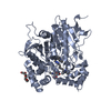
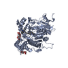
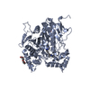
| Title | Nonaged form of Mouse Acetylcholinesterase inhibited by VX-Update | |||||||||
 Components Components | ACETYLCHOLINESTERASE | |||||||||
 Keywords Keywords | HYDROLASE / CHOLINESTERASE / METHYLPHOSPHONATE | |||||||||
| Function / homology |  Function and homology information Function and homology informationacetylcholine metabolic process / serine hydrolase activity / choline binding / acetylcholine catabolic process / acetylcholine binding / acetylcholinesterase / acetylcholine receptor signaling pathway / positive regulation of dendrite morphogenesis / osteoblast development / acetylcholinesterase activity ...acetylcholine metabolic process / serine hydrolase activity / choline binding / acetylcholine catabolic process / acetylcholine binding / acetylcholinesterase / acetylcholine receptor signaling pathway / positive regulation of dendrite morphogenesis / osteoblast development / acetylcholinesterase activity / choline metabolic process / positive regulation of axonogenesis / basement membrane / regulation of receptor recycling / laminin binding / side of membrane / synaptic cleft / synapse assembly / collagen binding / response to insulin / neuromuscular junction / receptor internalization / : / retina development in camera-type eye / nuclear envelope / presynaptic membrane / positive regulation of cold-induced thermogenesis / postsynaptic membrane / cell adhesion / endoplasmic reticulum lumen / axon / neuronal cell body / synapse / dendrite / perinuclear region of cytoplasm / Golgi apparatus / cell surface / protein homodimerization activity / extracellular space / identical protein binding / plasma membraneSimilarity search - Function | |||||||||
| Biological species |  MUS MUSCULUS (house mouse) MUS MUSCULUS (house mouse) | |||||||||
| Method | X-RAY DIFFRACTION / SYNCHROTRON / MOLECULAR REPLACEMENT / Resolution: 2.6 Å | |||||||||
 Authors Authors | Akfur, C. / Artursson, E. / Ekstrom, F. | |||||||||
 Citation Citation | Journal: To be Published Title: Methylphosphonate Adducts of Acetylcholinesterase Investigated by Time Correlated Single Photon Counting and X-Ray Crystallography Authors: Akfur, C. / Artursson, E. / Ekstrom, F. | |||||||||
| History |
|
- Structure visualization
| Structure viewer | Molecule: MolmilJmol/JSmol |
|---|
- Downloads & links
-Download
| PDBx/mmCIF format | 2y2u.cif.gz | 228.2 KB | Display | PDBx/mmCIF format |
|---|---|---|---|---|
| PDB format | pdb2y2u.ent.gz | 182.7 KB | Display | PDB format |
| PDBx/mmJSON format | 2y2u.json.gz | Tree view | PDBx/mmJSON format | |
| Others |  Other downloads Other downloads |
-Validation report
| Arichive directory | https://data.pdbj.org/pub/pdb/validation_reports/y2/2y2uftp://data.pdbj.org/pub/pdb/validation_reports/y2/2y2u | HTTPS FTP |
|---|
-Related structure data
| Related structure data |  2y2vC  1j06S C: citing same article ( S: Starting model for refinement |
|---|---|
| Similar structure data |


-Links
 PDBj
PDBj
- Assembly
| Deposited unit | 
| ||||||||
|---|---|---|---|---|---|---|---|---|---|
| 1 | 
| ||||||||
| 2 | 
| ||||||||
| Unit cell |
|
-Components
| #1: Protein | / ACHE Mass: 60340.047 Da / Num. of mol.: 2 / Fragment: CATALYTIC DOMAIN, RESIDUES 32-574 Source method: isolated from a genetically manipulated source Details: CATALYTIC SER203 PHOSPHONYLATED BY VX / Source: (gene. exp.) MUS MUSCULUS (house mouse) / Cell line (production host): HEK293F / Production host:  HOMO SAPIENS (human) / References: UniProt: P21836, acetylcholinesterase HOMO SAPIENS (human) / References: UniProt: P21836, acetylcholinesterase#2: Sugar | N-Acetylglucosamine  Type: D-saccharide, beta linking / Mass: 221.208 Da / Num. of mol.: 3 Type: D-saccharide, beta linking / Mass: 221.208 Da / Num. of mol.: 3Source method: isolated from a genetically manipulated source Formula: C8H15NO6 #3: Chemical | ChemComp-PEG / Diethylene glycol  Mass: 106.120 Da / Num. of mol.: 5 / Source method: obtained synthetically / Formula: C4H10O3 Mass: 106.120 Da / Num. of mol.: 5 / Source method: obtained synthetically / Formula: C4H10O3#4: Chemical | ChemComp-P33 / | Polyethylene glycol  Mass: 326.383 Da / Num. of mol.: 1 / Source method: obtained synthetically / Formula: C14H30O8 / Comment: precipitant*YM Mass: 326.383 Da / Num. of mol.: 1 / Source method: obtained synthetically / Formula: C14H30O8 / Comment: precipitant*YM#5: Water | ChemComp-HOH / | Water Mass: 18.015 Da / Num. of mol.: 357 / Source method: isolated from a natural source / Formula: H2O Mass: 18.015 Da / Num. of mol.: 357 / Source method: isolated from a natural source / Formula: H2ONonpolymer details | RESIDUE SVX REPRESENTS | |
|---|
-Experimental details
-Experiment
| Experiment | Method: X-RAY DIFFRACTION / Number of used crystals: 1 |
|---|
- Sample preparation
| Crystal | Density Matthews: 2.39 Å3/Da / Density % sol: 48.6 % / Description: NONE |
|---|---|
| Crystal grow | pH: 7.9 / Details: 26-30% (V/V) PEG 750MME, 0.1 M HEPES PH 7.0. |
-Data collection
| Diffraction | Mean temperature: 100 K |
|---|---|
| Diffraction source | Source: SYNCHROTRON / Site: MAX II  / Beamline: I911-3 / Wavelength: 1 / Beamline: I911-3 / Wavelength: 1 |
| Detector | Type: MARMOSAIC 225 mm CCD / Detector: CCD / Date: Jun 2, 2010 / Details: MIRRORS |
| Radiation | Protocol: SINGLE WAVELENGTH / Monochromatic (M) / Laue (L): M / Scattering type: x-ray |
| Radiation wavelength | Wavelength: 1 Å / Relative weight: 1 |
| Reflection | Resolution: 2.6→29.03 Å / Num. obs: 61880 / % possible obs: 99.9 % / Observed criterion σ(I): 3.7 / Redundancy: 7.3 % / Rmerge(I) obs: 0.08 / Net I/σ(I): 16.6 |
| Reflection shell | Resolution: 2.6→2.74 Å / Redundancy: 7.1 % / Rmerge(I) obs: 0.54 / Mean I/σ(I) obs: 3.7 / % possible all: 99.9 |
- Processing
| Software |
| ||||||||||||||||||||||||||||||||||||||||||||||||||||||||||||||||||||||
|---|---|---|---|---|---|---|---|---|---|---|---|---|---|---|---|---|---|---|---|---|---|---|---|---|---|---|---|---|---|---|---|---|---|---|---|---|---|---|---|---|---|---|---|---|---|---|---|---|---|---|---|---|---|---|---|---|---|---|---|---|---|---|---|---|---|---|---|---|---|---|---|
| Refinement | Method to determine structure: MOLECULAR REPLACEMENT Starting model: PDB ENTRY 1J06 Resolution: 2.6→28.762 Å / SU ML: 0.73 / σ(F): 1.33 / Phase error: 22.16 / Stereochemistry target values: ML
| ||||||||||||||||||||||||||||||||||||||||||||||||||||||||||||||||||||||
| Solvent computation | Shrinkage radii: 0.95 Å / VDW probe radii: 1.2 Å / Solvent model: FLAT BULK SOLVENT MODEL / Bsol: 63.076 Å2 / ksol: 0.346 e/Å3 | ||||||||||||||||||||||||||||||||||||||||||||||||||||||||||||||||||||||
| Displacement parameters |
| ||||||||||||||||||||||||||||||||||||||||||||||||||||||||||||||||||||||
| Refinement step | Cycle: LAST / Resolution: 2.6→28.762 Å
| ||||||||||||||||||||||||||||||||||||||||||||||||||||||||||||||||||||||
| Refine LS restraints |
| ||||||||||||||||||||||||||||||||||||||||||||||||||||||||||||||||||||||
| LS refinement shell |
|
