 Movie
Movie Controller

[English] 日本語
 Yorodumi
- PDB-2xuo: CRYSTAL STRUCTURE OF MACHE-Y337A mutant in complex with soaked TZ... -
+ Open data

- Basic information
| Entry | Database: PDB / ID: 2xuo | ||||||
|---|---|---|---|---|---|---|---|
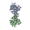
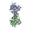
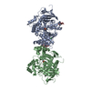
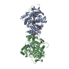
| Title | CRYSTAL STRUCTURE OF MACHE-Y337A mutant in complex with soaked TZ2PA6 ANTI inhibitor | ||||||
 Components Components | ACETYLCHOLINESTERASE | ||||||
 Keywords Keywords | HYDROLASE / HYDROLASE FOLD / CLICK CHEMISTRY | ||||||
| Function / homology |  Function and homology information Function and homology informationacetylcholine metabolic process / serine hydrolase activity / choline metabolic process / choline binding / acetylcholine catabolic process / acetylcholine binding / acetylcholinesterase / cholinesterase activity / acetylcholine receptor signaling pathway / positive regulation of dendrite morphogenesis ...acetylcholine metabolic process / serine hydrolase activity / choline metabolic process / choline binding / acetylcholine catabolic process / acetylcholine binding / acetylcholinesterase / cholinesterase activity / acetylcholine receptor signaling pathway / positive regulation of dendrite morphogenesis / osteoblast development / acetylcholinesterase activity / positive regulation of axonogenesis / basement membrane / regulation of receptor recycling / laminin binding / side of membrane / synaptic cleft / synapse assembly / collagen binding / response to insulin / neuromuscular junction / receptor internalization / : / retina development in camera-type eye / presynaptic membrane / nuclear envelope / positive regulation of cold-induced thermogenesis / postsynaptic membrane / hydrolase activity / cell adhesion / axon / endoplasmic reticulum lumen / neuronal cell body / synapse / dendrite / perinuclear region of cytoplasm / Golgi apparatus / cell surface / protein homodimerization activity / extracellular space / extracellular region / membrane / identical protein binding / plasma membraneSimilarity search - Function | ||||||
| Biological species |  MUS MUSCULUS (house mouse) MUS MUSCULUS (house mouse) | ||||||
| Method | X-RAY DIFFRACTION / SYNCHROTRON / MOLECULAR REPLACEMENT / Resolution: 2.8 Å | ||||||
 Authors Authors | Bourne, Y. / Radic, Z. / Taylor, P. / Marchot, P. | ||||||
 Citation Citation | Journal: J.Am.Chem.Soc. / Year: 2010 Title: Conformational Remodeling of Femtomolar Inhibitor-Acetylcholinesterase Complexes in the Crystalline State Authors: Bourne, Y. / Radic, Z. / Taylor, P. / Marchot, P. | ||||||
| History |
|
- Structure visualization
| Structure viewer | Molecule: MolmilJmol/JSmol |
|---|
- Downloads & links
-Download
| PDBx/mmCIF format | 2xuo.cif.gz | 425.4 KB | Display | PDBx/mmCIF format |
|---|---|---|---|---|
| PDB format | pdb2xuo.ent.gz | 352.2 KB | Display | PDB format |
| PDBx/mmJSON format | 2xuo.json.gz | Tree view | PDBx/mmJSON format | |
| Others |  Other downloads Other downloads |
-Validation report
| Arichive directory | https://data.pdbj.org/pub/pdb/validation_reports/xu/2xuoftp://data.pdbj.org/pub/pdb/validation_reports/xu/2xuo | HTTPS FTP |
|---|
-Related structure data
| Related structure data |  2xudC  2xufC  2xugC  2xuhC  2xuiC  2xujC  2xukC  2xupC  2xuqC  1j06S C: citing same article ( S: Starting model for refinement |
|---|---|
| Similar structure data |


-Links
 PDBj
PDBj
- Assembly
| Deposited unit | 
| |||||||||||||||||||||||||||||||||||||||||||||||||||||||||||||||||||||||||||||||||||||||||||||||||||||||||||||||||||||||||||||||||||||||||||
|---|---|---|---|---|---|---|---|---|---|---|---|---|---|---|---|---|---|---|---|---|---|---|---|---|---|---|---|---|---|---|---|---|---|---|---|---|---|---|---|---|---|---|---|---|---|---|---|---|---|---|---|---|---|---|---|---|---|---|---|---|---|---|---|---|---|---|---|---|---|---|---|---|---|---|---|---|---|---|---|---|---|---|---|---|---|---|---|---|---|---|---|---|---|---|---|---|---|---|---|---|---|---|---|---|---|---|---|---|---|---|---|---|---|---|---|---|---|---|---|---|---|---|---|---|---|---|---|---|---|---|---|---|---|---|---|---|---|---|---|---|
| 1 | 
| |||||||||||||||||||||||||||||||||||||||||||||||||||||||||||||||||||||||||||||||||||||||||||||||||||||||||||||||||||||||||||||||||||||||||||
| Unit cell |
| |||||||||||||||||||||||||||||||||||||||||||||||||||||||||||||||||||||||||||||||||||||||||||||||||||||||||||||||||||||||||||||||||||||||||||
| Noncrystallographic symmetry (NCS) | NCS domain:
NCS domain segments: Ens-ID: 1 / Refine code: 5
NCS oper:
|
-Components
| #1: Protein | / ACHE Mass: 59672.395 Da / Num. of mol.: 2 / Fragment: CATALYTIC DOMAIN, RESIDUES 32-574 / Mutation: YES Source method: isolated from a genetically manipulated source Source: (gene. exp.) MUS MUSCULUS (house mouse) / Strain: BLACK6CBA CROSS F1 / Description: LAMBDA-FIX CDNA, GENOMIC DNA / Organ: BRAIN (CDNA) / Plasmid: LAMBDA-ZAP / Cell line (production host): HEK293 / Production host:  HOMO SAPIENS (human) / References: UniProt: P21836, acetylcholinesterase HOMO SAPIENS (human) / References: UniProt: P21836, acetylcholinesterase#2: Chemical | ChemComp-P6G / | Polyethylene glycol  Mass: 282.331 Da / Num. of mol.: 1 / Source method: obtained synthetically / Formula: C12H26O7 / Comment: precipitant*YM Mass: 282.331 Da / Num. of mol.: 1 / Source method: obtained synthetically / Formula: C12H26O7 / Comment: precipitant*YM#3: Chemical |   Mass: 661.860 Da / Num. of mol.: 2 / Source method: obtained synthetically / Formula: C42H45N8 Mass: 661.860 Da / Num. of mol.: 2 / Source method: obtained synthetically / Formula: C42H45N8#4: Water | ChemComp-HOH / | Water Mass: 18.015 Da / Num. of mol.: 37 / Source method: isolated from a natural source / Formula: H2O Mass: 18.015 Da / Num. of mol.: 37 / Source method: isolated from a natural source / Formula: H2OCompound details | ENGINEERED | |
|---|
-Experimental details
-Experiment
| Experiment | Method: X-RAY DIFFRACTION / Number of used crystals: 1 |
|---|
- Sample preparation
| Crystal | Density Matthews: 4.08 Å3/Da / Density % sol: 69.6 % / Description: NONE |
|---|---|
| Crystal grow | pH: 6.5 Details: PROTEIN WAS CRYSTALLIZED FROM PEG-600 25-35% (V/V) IN 50-100 MM HEPES, PH 6.0-7.0, OR WITH PEG-550 MME 30% (V/V) IN 50 MM NA ACETATE, PH 7.5 |
-Data collection
| Diffraction | Mean temperature: 100 K |
|---|---|
| Diffraction source | Source: SYNCHROTRON / Site: ESRF  / Beamline: ID14-3 / Wavelength: 0.931 / Beamline: ID14-3 / Wavelength: 0.931 |
| Detector | Type: ADSC CCD / Detector: CCD / Date: Mar 22, 2006 |
| Radiation | Protocol: SINGLE WAVELENGTH / Monochromatic (M) / Laue (L): M / Scattering type: x-ray |
| Radiation wavelength | Wavelength: 0.931 Å / Relative weight: 1 |
| Reflection | Resolution: 2.8→20 Å / Num. obs: 49788 / % possible obs: 99.8 % / Observed criterion σ(I): 2 / Redundancy: 5.2 % / Biso Wilson estimate: 72 Å2 / Rmerge(I) obs: 0.09 / Net I/σ(I): 16.6 |
| Reflection shell | Resolution: 2.8→2.95 Å / Redundancy: 5.1 % / Rmerge(I) obs: 0.54 / Mean I/σ(I) obs: 3.6 / % possible all: 99.8 |
- Processing
| Software |
| ||||||||||||||||||||||||||||||||||||||||||||||||||||||||||||||||||||||||||||||||||||||||||||||||||||||||||||||||||||||||||||||||||||||||||||||||||||||||||||||||||||||||||||||||||||||
|---|---|---|---|---|---|---|---|---|---|---|---|---|---|---|---|---|---|---|---|---|---|---|---|---|---|---|---|---|---|---|---|---|---|---|---|---|---|---|---|---|---|---|---|---|---|---|---|---|---|---|---|---|---|---|---|---|---|---|---|---|---|---|---|---|---|---|---|---|---|---|---|---|---|---|---|---|---|---|---|---|---|---|---|---|---|---|---|---|---|---|---|---|---|---|---|---|---|---|---|---|---|---|---|---|---|---|---|---|---|---|---|---|---|---|---|---|---|---|---|---|---|---|---|---|---|---|---|---|---|---|---|---|---|---|---|---|---|---|---|---|---|---|---|---|---|---|---|---|---|---|---|---|---|---|---|---|---|---|---|---|---|---|---|---|---|---|---|---|---|---|---|---|---|---|---|---|---|---|---|---|---|---|---|
| Refinement | Method to determine structure: MOLECULAR REPLACEMENT Starting model: PDB ENTRY 1J06 Resolution: 2.8→20 Å / Cor.coef. Fo:Fc: 0.946 / Cor.coef. Fo:Fc free: 0.916 / SU B: 26.145 / SU ML: 0.232 / Cross valid method: THROUGHOUT / ESU R: 0.429 / ESU R Free: 0.291 / Stereochemistry target values: MAXIMUM LIKELIHOOD / Details: HYDROGENS HAVE BEEN ADDED IN THE RIDING POSITIONS.
| ||||||||||||||||||||||||||||||||||||||||||||||||||||||||||||||||||||||||||||||||||||||||||||||||||||||||||||||||||||||||||||||||||||||||||||||||||||||||||||||||||||||||||||||||||||||
| Solvent computation | Ion probe radii: 0.8 Å / Shrinkage radii: 0.8 Å / VDW probe radii: 1.4 Å / Solvent model: MASK | ||||||||||||||||||||||||||||||||||||||||||||||||||||||||||||||||||||||||||||||||||||||||||||||||||||||||||||||||||||||||||||||||||||||||||||||||||||||||||||||||||||||||||||||||||||||
| Displacement parameters | Biso mean: 59.252 Å2
| ||||||||||||||||||||||||||||||||||||||||||||||||||||||||||||||||||||||||||||||||||||||||||||||||||||||||||||||||||||||||||||||||||||||||||||||||||||||||||||||||||||||||||||||||||||||
| Refinement step | Cycle: LAST / Resolution: 2.8→20 Å
| ||||||||||||||||||||||||||||||||||||||||||||||||||||||||||||||||||||||||||||||||||||||||||||||||||||||||||||||||||||||||||||||||||||||||||||||||||||||||||||||||||||||||||||||||||||||
| Refine LS restraints |
|