 Movie
Movie Controller

[English] 日本語
 Yorodumi
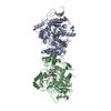
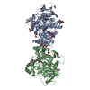
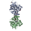
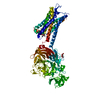
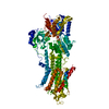
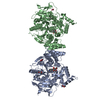
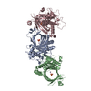
- PDB-3bix: Crystal structure of the extracellular esterase domain of Neuroligin-1 -
+ Open data

- Basic information
| Entry | Database: PDB / ID: 3bix | ||||||
|---|---|---|---|---|---|---|---|
| Title | Crystal structure of the extracellular esterase domain of Neuroligin-1 | ||||||
 Components Components | Neuroligin-1 | ||||||
 Keywords Keywords | CELL ADHESION / esterase domain / alpha-beta hydrolase / Cell junction / Glycoprotein / Membrane / Postsynaptic cell membrane / Synapse / Transmembrane | ||||||
| Function / homology |  Function and homology information Function and homology informationneurexin clustering involved in presynaptic membrane assembly / regulation of presynapse organization / positive regulation of presynaptic active zone assembly / cytoskeletal matrix organization at active zone / cell-cell adhesion involved in synapse maturation / positive regulation of circadian sleep/wake cycle, wakefulness / retrograde trans-synaptic signaling by trans-synaptic protein complex / protein complex involved in cell-cell adhesion / positive regulation of neuromuscular synaptic transmission / terminal button organization ...neurexin clustering involved in presynaptic membrane assembly / regulation of presynapse organization / positive regulation of presynaptic active zone assembly / cytoskeletal matrix organization at active zone / cell-cell adhesion involved in synapse maturation / positive regulation of circadian sleep/wake cycle, wakefulness / retrograde trans-synaptic signaling by trans-synaptic protein complex / protein complex involved in cell-cell adhesion / positive regulation of neuromuscular synaptic transmission / terminal button organization / neuron to neuron synapse / positive regulation of synaptic vesicle exocytosis / excitatory synapse assembly / postsynaptic density protein 95 clustering / postsynaptic specialization assembly / negative regulation of dendritic spine morphogenesis / postsynaptic membrane assembly / neuronal ion channel clustering / positive regulation of synaptic vesicle clustering / presynaptic membrane assembly / synapse maturation / maintenance of synapse structure / Neurexins and neuroligins / synaptic vesicle targeting / synaptic membrane adhesion / synaptic vesicle clustering / inhibitory synapse / receptor localization to synapse / neuron cell-cell adhesion / regulation of respiratory gaseous exchange by nervous system process / neurexin family protein binding / filopodium tip / protein localization to synapse / NMDA glutamate receptor clustering / neuron projection arborization / positive regulation of synaptic vesicle endocytosis / calcium-dependent cell-cell adhesion via plasma membrane cell adhesion molecules / regulation of AMPA receptor activity / AMPA glutamate receptor clustering / postsynaptic specialization membrane / positive regulation of synapse assembly / positive regulation of ruffle assembly / positive regulation of filopodium assembly / positive regulation of protein localization to synapse / positive regulation of intracellular signal transduction / heterophilic cell-cell adhesion via plasma membrane cell adhesion molecules / synaptic vesicle transport / regulation of neuron differentiation / positive regulation of dendritic spine development / regulation of NMDA receptor activity / positive regulation of excitatory postsynaptic potential / excitatory synapse / synaptic vesicle endocytosis / protein targeting / GABA-ergic synapse / synaptic cleft / synapse assembly / cellular response to calcium ion / cell adhesion molecule binding / positive regulation of synaptic transmission, glutamatergic / neuron projection morphogenesis / dendritic shaft / long-term synaptic potentiation / PDZ domain binding / positive regulation of synaptic transmission, GABAergic / synapse organization / modulation of chemical synaptic transmission / neuromuscular junction / establishment of protein localization / positive regulation of neuron projection development / neuron projection development / rhythmic process / presynapse / signaling receptor activity / nervous system development / chemical synaptic transmission / scaffold protein binding / dendritic spine / postsynaptic density / receptor complex / external side of plasma membrane / dendrite / synapse / glutamatergic synapse / protein-containing complex binding / Golgi apparatus / cell surface / identical protein binding / plasma membraneSimilarity search - Function | ||||||
| Biological species |  Rattus norvegicus (Norway rat) Rattus norvegicus (Norway rat) | ||||||
| Method | X-RAY DIFFRACTION / SYNCHROTRON / MOLECULAR REPLACEMENT / molecular replacement / Resolution: 1.8 Å | ||||||
 Authors Authors | Arac, D. / Boucard, A.A. / Ozkan, E. / Strop, P. / Newell, E. / Sudhof, T.C. / Brunger, A.T. | ||||||
 Citation Citation | Journal: Neuron / Year: 2007 Title: Structures of Neuroligin-1 and the Neuroligin-1/Neurexin-1beta Complex Reveal Specific Protein-Protein and Protein-Ca(2+) Interactions. Authors: Arac, D. / Boucard, A.A. / Ozkan, E. / Strop, P. / Newell, E. / Sudhof, T.C. / Brunger, A.T. | ||||||
| History |
|
- Structure visualization
| Structure viewer | Molecule: MolmilJmol/JSmol |
|---|
- Downloads & links
-Download
| PDBx/mmCIF format | 3bix.cif.gz | 480.8 KB | Display | PDBx/mmCIF format |
|---|---|---|---|---|
| PDB format | pdb3bix.ent.gz | 394.3 KB | Display | PDB format |
| PDBx/mmJSON format | 3bix.json.gz | Tree view | PDBx/mmJSON format | |
| Others |  Other downloads Other downloads |
-Validation report
| Arichive directory | https://data.pdbj.org/pub/pdb/validation_reports/bi/3bixftp://data.pdbj.org/pub/pdb/validation_reports/bi/3bix | HTTPS FTP |
|---|
-Related structure data


-Links
 PDBj
PDBj
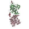
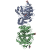
- Assembly
| Deposited unit | 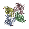
| ||||||||
|---|---|---|---|---|---|---|---|---|---|
| 1 | 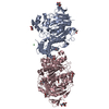
| ||||||||
| 2 | 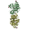
| ||||||||
| Unit cell |
|
-Components
| #1: Protein | / Neuroligin I Mass: 64228.961 Da / Num. of mol.: 4 / Fragment: extracellular esterase domain Source method: isolated from a genetically manipulated source Source: (gene. exp.) Rattus norvegicus (Norway rat) / Gene: Nlgn1 / Plasmid: pAcGP67A / Production host:  Trichoplusia ni (cabbage looper) / References: UniProt: Q62765 Trichoplusia ni (cabbage looper) / References: UniProt: Q62765#2: Sugar | ChemComp-NAG / N-Acetylglucosamine  Type: D-saccharide, beta linking / Mass: 221.208 Da / Num. of mol.: 8 Type: D-saccharide, beta linking / Mass: 221.208 Da / Num. of mol.: 8Source method: isolated from a genetically manipulated source Formula: C8H15NO6 #3: Chemical | Nickel  Mass: 58.693 Da / Num. of mol.: 2 / Source method: obtained synthetically / Formula: Ni Mass: 58.693 Da / Num. of mol.: 2 / Source method: obtained synthetically / Formula: Ni#4: Chemical | ChemComp-EDO / Ethylene glycol  Mass: 62.068 Da / Num. of mol.: 14 / Source method: obtained synthetically / Formula: C2H6O2 Mass: 62.068 Da / Num. of mol.: 14 / Source method: obtained synthetically / Formula: C2H6O2#5: Water | ChemComp-HOH / | Water Mass: 18.015 Da / Num. of mol.: 1965 / Source method: isolated from a natural source / Formula: H2O Mass: 18.015 Da / Num. of mol.: 1965 / Source method: isolated from a natural source / Formula: H2O |
|---|
-Experimental details
-Experiment
| Experiment | Method: X-RAY DIFFRACTION / Number of used crystals: 1 |
|---|
- Sample preparation
| Crystal | Density Matthews: 3.66 Å3/Da / Density % sol: 66.44 % |
|---|---|
| Crystal grow | Temperature: 293 K / Method: vapor diffusion, hanging drop / pH: 8.7 Details: 1.2 M tri-Sodium citrate, 0.1 M Tris, pH 8.7, VAPOR DIFFUSION, HANGING DROP, temperature 293K |
-Data collection
| Diffraction | Mean temperature: 130 K | |||||||||||||||||||||||||||||||||||||||||||||||||||||||||||||||||||||||||||||
|---|---|---|---|---|---|---|---|---|---|---|---|---|---|---|---|---|---|---|---|---|---|---|---|---|---|---|---|---|---|---|---|---|---|---|---|---|---|---|---|---|---|---|---|---|---|---|---|---|---|---|---|---|---|---|---|---|---|---|---|---|---|---|---|---|---|---|---|---|---|---|---|---|---|---|---|---|---|---|
| Diffraction source | Source: SYNCHROTRON / Site: ALS  / Beamline: 8.2.1 / Wavelength: 1 Å / Beamline: 8.2.1 / Wavelength: 1 Å | |||||||||||||||||||||||||||||||||||||||||||||||||||||||||||||||||||||||||||||
| Detector | Type: ADSC QUANTUM 315 / Detector: CCD / Date: Feb 15, 2007 | |||||||||||||||||||||||||||||||||||||||||||||||||||||||||||||||||||||||||||||
| Radiation | Monochromator: Double crystal, Si(111) / Protocol: SINGLE WAVELENGTH / Monochromatic (M) / Laue (L): M / Scattering type: x-ray | |||||||||||||||||||||||||||||||||||||||||||||||||||||||||||||||||||||||||||||
| Radiation wavelength | Wavelength: 1 Å / Relative weight: 1 | |||||||||||||||||||||||||||||||||||||||||||||||||||||||||||||||||||||||||||||
| Reflection | Resolution: 1.75→50 Å / Num. obs: 360486 / % possible obs: 98.3 % / Redundancy: 2.7 % / Rmerge(I) obs: 0.077 / Χ2: 0.992 / Net I/σ(I): 10.2 | |||||||||||||||||||||||||||||||||||||||||||||||||||||||||||||||||||||||||||||
| Reflection shell |
|
-Phasing
| Phasing | Method: molecular replacement |
|---|
- Processing
| Software |
| ||||||||||||||||||||||||||||||||||||
|---|---|---|---|---|---|---|---|---|---|---|---|---|---|---|---|---|---|---|---|---|---|---|---|---|---|---|---|---|---|---|---|---|---|---|---|---|---|
| Refinement | Method to determine structure: MOLECULAR REPLACEMENT / Resolution: 1.8→45.88 Å / Rfactor Rfree error: 0.002 / FOM work R set: 0.866 / Data cutoff high absF: 1687471.75 / Data cutoff low absF: 0 / Isotropic thermal model: RESTRAINED / Cross valid method: THROUGHOUT / σ(F): 0 / Details: BULK SOLVENT MODEL USED
| ||||||||||||||||||||||||||||||||||||
| Solvent computation | Solvent model: FLAT MODEL / Bsol: 54.314 Å2 / ksol: 0.4 e/Å3 | ||||||||||||||||||||||||||||||||||||
| Displacement parameters | Biso mean: 25.1 Å2
| ||||||||||||||||||||||||||||||||||||
| Refine analyze |
| ||||||||||||||||||||||||||||||||||||
| Refinement step | Cycle: LAST / Resolution: 1.8→45.88 Å
| ||||||||||||||||||||||||||||||||||||
| Refine LS restraints |
| ||||||||||||||||||||||||||||||||||||
| LS refinement shell | Resolution: 1.8→1.91 Å / Rfactor Rfree error: 0.006 / Total num. of bins used: 6
| ||||||||||||||||||||||||||||||||||||
| Xplor file |
|
