 Movie
Movie Controller

[English] 日本語
 Yorodumi
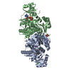
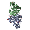
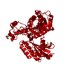
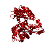
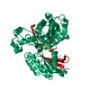
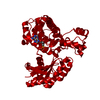
- PDB-1d8h: X-RAY CRYSTAL STRUCTURE OF YEAST RNA TRIPHOSPHATASE IN COMPLEX WI... -
+ Open data

- Basic information
| Entry | Database: PDB / ID: 1d8h | ||||||
|---|---|---|---|---|---|---|---|
| Title | X-RAY CRYSTAL STRUCTURE OF YEAST RNA TRIPHOSPHATASE IN COMPLEX WITH SULFATE AND MANGANESE IONS. | ||||||
 Components Components | mRNA TRIPHOSPHATASE CET1 | ||||||
 Keywords Keywords |  HYDROLASE / RNA TRIPHOSPHATASE / BETA SUBUNIT / POLYNUCLEOTIDE 5'-TRIPHOSPHATASE / mRNA PROCESSING / mRNA CAPPING / NUCLEAR PROTEIN BETA BARREL / CATALYTIC DOMAIN / DIMER / MANGANESE-SULFATE COMPLEX HYDROLASE / RNA TRIPHOSPHATASE / BETA SUBUNIT / POLYNUCLEOTIDE 5'-TRIPHOSPHATASE / mRNA PROCESSING / mRNA CAPPING / NUCLEAR PROTEIN BETA BARREL / CATALYTIC DOMAIN / DIMER / MANGANESE-SULFATE COMPLEX | ||||||
| Function / homology |  Function and homology information Function and homology informationpolynucleotide 5' dephosphorylation / mRNA capping enzyme complex / mRNA 5'-triphosphate monophosphatase activity / mRNA 5'-phosphatase / polynucleotide 5'-phosphatase activity / 7-methylguanosine mRNA capping / positive regulation of transcription elongation by RNA polymerase II / positive regulation of protein localization to nucleusSimilarity search - Function | ||||||
| Biological species |  Saccharomyces cerevisiae (brewer's yeast) Saccharomyces cerevisiae (brewer's yeast) | ||||||
| Method | X-RAY DIFFRACTION / SYNCHROTRON / Resolution: 2 Å | ||||||
 Authors Authors | Lima, C.D. / Wang, L.K. / Shuman, S. | ||||||
 Citation Citation | Journal: Cell(Cambridge,Mass.) / Year: 1999 Title: Structure and mechanism of yeast RNA triphosphatase: an essential component of the mRNA capping apparatus. Authors: Lima, C.D. / Wang, L.K. / Shuman, S. | ||||||
| History |
|
- Structure visualization
| Structure viewer | Molecule: MolmilJmol/JSmol |
|---|
- Downloads & links
-Download
| PDBx/mmCIF format | 1d8h.cif.gz | 212.6 KB | Display | PDBx/mmCIF format |
|---|---|---|---|---|
| PDB format | pdb1d8h.ent.gz | 161.7 KB | Display | PDB format |
| PDBx/mmJSON format | 1d8h.json.gz | Tree view | PDBx/mmJSON format | |
| Others |  Other downloads Other downloads |
-Validation report
| Arichive directory | https://data.pdbj.org/pub/pdb/validation_reports/d8/1d8hftp://data.pdbj.org/pub/pdb/validation_reports/d8/1d8h | HTTPS FTP |
|---|
-Related structure data


-Links
 PDBj
PDBj
- Assembly
| Deposited unit | 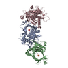
| ||||||||
|---|---|---|---|---|---|---|---|---|---|
| 1 | 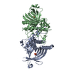
| ||||||||
| 2 | 
| ||||||||
| Unit cell |
|
-Components
| #1: Protein | Mass: 35691.465 Da / Num. of mol.: 3 Source method: isolated from a genetically manipulated source Source: (gene. exp.) Saccharomyces cerevisiae (brewer's yeast)Plasmid details: T7 PROMOTER / Plasmid: PET16B / Species (production host): Escherichia coli / Production host:  Escherichia coli BL21(DE3) (bacteria) / Strain (production host): BL21 (DE3) / References: UniProt: O13297, polynucleotide 5'-phosphatase Escherichia coli BL21(DE3) (bacteria) / Strain (production host): BL21 (DE3) / References: UniProt: O13297, polynucleotide 5'-phosphatase#2: Chemical |   Mass: 54.938 Da / Num. of mol.: 3 / Source method: obtained synthetically / Formula: Mn Mass: 54.938 Da / Num. of mol.: 3 / Source method: obtained synthetically / Formula: Mn#3: Chemical | Sulfate  Mass: 96.063 Da / Num. of mol.: 3 / Source method: obtained synthetically / Formula: SO4 Mass: 96.063 Da / Num. of mol.: 3 / Source method: obtained synthetically / Formula: SO4#4: Water | ChemComp-HOH / | Water Mass: 18.015 Da / Num. of mol.: 669 / Source method: isolated from a natural source / Formula: H2O Mass: 18.015 Da / Num. of mol.: 669 / Source method: isolated from a natural source / Formula: H2OCompound details | RNA triphosphatase is an essential mRNA processing enzyme that catalyzes the first step in 5' cap ...RNA triphosphatase is an essential mRNA processing enzyme that catalyzes the first step in 5' cap formation. The 2.05 A crystal structure of yeast RNA triphosphatase Cet1p reveals a novel active site fold whereby an 8-strand beta barrel forms a topologically closed triphosphatase tunnel. Interactions of a sulfate bound in the center of the tunnel with a divalent cation and basic amino acids projecting into the tunnel suggest a catalytic mechanism that is supported by mutational data. Discrete surface domains are responsible for Cet1p homodimerization and Cet1p-binding to the guanylyltransferase component of the yeast capping apparatus. The structure and mechanism of the fungal RNA triphosphatases are completely different from those of the mammalian mRNA capping enzymes. | Sequence details | THE EXPRESSION CONSTRUCTS USED TO PRODUCE THE RECOMBINANT PROTEIN INCLUDED AN N-TERMINAL HIS TAG ...THE EXPRESSION | |
|---|
-Experimental details
-Experiment
| Experiment | Method: X-RAY DIFFRACTION / Number of used crystals: 1 |
|---|
- Sample preparation
| Crystal | Density Matthews: 2.61 Å3/Da / Density % sol: 52.9 % | |||||||||||||||||||||||||
|---|---|---|---|---|---|---|---|---|---|---|---|---|---|---|---|---|---|---|---|---|---|---|---|---|---|---|
| Crystal grow | Temperature: 295 K / Method: vapor diffusion, hanging drop / pH: 6.34 Details: 0.1M MES, 38% SATURATED (NH4)2SO4, 5MM DTT STABILIZED IN 2.5M AMSO4 + BUFFER, 200MM MNCL2 IN STABILIZATION CONDITION FOR MANGANESE COMPLEX, pH 6.34, VAPOR DIFFUSION, HANGING DROP, temperature 295K | |||||||||||||||||||||||||
| Crystal grow | *PLUS Temperature: 22 ℃Details: drop solution was mixed with an equal volume of reservoir solution | |||||||||||||||||||||||||
| Components of the solutions | *PLUS
|
-Data collection
| Diffraction | Mean temperature: 100 K |
|---|---|
| Diffraction source | Source: SYNCHROTRON / Site: NSLS  / Beamline: X4A / Wavelength: 1.2141 / Beamline: X4A / Wavelength: 1.2141 |
| Detector | Type: ADSC QUANTUM 4 / Detector: CCD / Date: Aug 8, 1999 |
| Radiation | Protocol: SINGLE WAVELENGTH / Monochromatic (M) / Laue (L): M / Scattering type: x-ray |
| Radiation wavelength | Wavelength: 1.2141 Å / Relative weight: 1 |
| Reflection | Resolution: 2→25 Å / Num. all: 72277 / Num. obs: 63710 / % possible obs: 88.1 % / Observed criterion σ(F): 0 / Observed criterion σ(I): 0 / Redundancy: 2.3 % / Biso Wilson estimate: 22.199 Å2 / Rmerge(I) obs: 0.066 / Net I/σ(I): 14.6 |
| Reflection shell | Resolution: 2→2.07 Å / Redundancy: 1.5 % / Rmerge(I) obs: 0.261 / % possible all: 72.2 |
| Reflection shell | *PLUS % possible obs: 72.2 % |
- Processing
| Software |
| |||||||||||||||||||||||||||||||||||||||||||||||||||||||||||||||
|---|---|---|---|---|---|---|---|---|---|---|---|---|---|---|---|---|---|---|---|---|---|---|---|---|---|---|---|---|---|---|---|---|---|---|---|---|---|---|---|---|---|---|---|---|---|---|---|---|---|---|---|---|---|---|---|---|---|---|---|---|---|---|---|---|
| Refinement | Resolution: 2→25 Å / σ(F): 0 / σ(I): 0 / Stereochemistry target values: ENGH & HUBER Details: RESTRAINED MAXIMUM LIKELIHOOD FOR FS CONJUGATE DIRECTION EXPERIMENTAL SIGMAS USED FOR WEIGHTING
| |||||||||||||||||||||||||||||||||||||||||||||||||||||||||||||||
| Refinement step | Cycle: LAST / Resolution: 2→25 Å
| |||||||||||||||||||||||||||||||||||||||||||||||||||||||||||||||
| Refine LS restraints |
|
