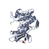
 Movie
Movie Controller

[English] 日本語
 Yorodumi
- PDB-5wno: Crystal structure of C. elegans LET-23 kinase domain complexed wi... -
+ Open data

- Basic information
| Entry | Database: PDB / ID: 5wno | ||||||||||||
|---|---|---|---|---|---|---|---|---|---|---|---|---|---|
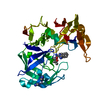
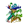
| Title | Crystal structure of C. elegans LET-23 kinase domain complexed with AMP-PNP | ||||||||||||
 Components Components | Receptor tyrosine-protein kinase let-23 | ||||||||||||
 Keywords Keywords |  TRANSFERASE / Receptor tyrosine-protein kinase / dimerization / inactive conformation / cell signaling / Let-23 / C. elegans TRANSFERASE / Receptor tyrosine-protein kinase / dimerization / inactive conformation / cell signaling / Let-23 / C. elegans | ||||||||||||
| Function / homology |  Function and homology information Function and homology informationvulval cell fate specification / SHC1 events in ERBB2 signaling / Nuclear signaling by ERBB4 / Downregulation of ERBB4 signaling / GAB1 signalosome / : / Sema4D induced cell migration and growth-cone collapse / Signal transduction by L1 / ERBB2 Regulates Cell Motility / ERBB2 Activates PTK6 Signaling ...vulval cell fate specification / SHC1 events in ERBB2 signaling / Nuclear signaling by ERBB4 / Downregulation of ERBB4 signaling / GAB1 signalosome / : / Sema4D induced cell migration and growth-cone collapse / Signal transduction by L1 / ERBB2 Regulates Cell Motility / ERBB2 Activates PTK6 Signaling / Signaling by EGFR / Signaling by ERBB2 / Downregulation of ERBB2 signaling / Extra-nuclear estrogen signaling / positive regulation of vulval development / PIP3 activates AKT signaling / GRB2 events in EGFR signaling / SHC1 events in EGFR signaling / EGFR downregulation / PI3K events in ERBB2 signaling / EGFR Transactivation by Gastrin / RAF/MAP kinase cascade / PI5P, PP2A and IER3 Regulate PI3K/AKT Signaling / Cargo recognition for clathrin-mediated endocytosis / Clathrin-mediated endocytosis / nematode larval development / ovulation / multicellular organism development / male genitalia development / sleep / positive regulation of kinase activity / epidermal growth factor receptor activity / plasma membrane => GO:0005886 / lateral plasma membrane / transmembrane receptor protein tyrosine kinase activity / basal plasma membrane / determination of adult lifespan / cell surface receptor protein tyrosine kinase signaling pathway / epidermal growth factor receptor signaling pathway / receptor protein-tyrosine kinase / cell-cell junction / positive regulation of proteasomal ubiquitin-dependent protein catabolic process / basolateral plasma membrane / receptor complex / apical plasma membrane / positive regulation of cell population proliferation / regulation of DNA-templated transcription / ATP bindingSimilarity search - Function | ||||||||||||
| Biological species |  Caenorhabditis elegans (invertebrata) Caenorhabditis elegans (invertebrata) | ||||||||||||
| Method | X-RAY DIFFRACTION / SYNCHROTRON / MOLECULAR REPLACEMENT / Resolution: 2.386 Å | ||||||||||||
 Authors Authors | Liu, L. / Thaker, T.M. / Jura, N. | ||||||||||||
| Funding support |  United States, 3items United States, 3items
| ||||||||||||
 Citation Citation | Journal: Structure / Year: 2018 Title: Regulation of Kinase Activity in the Caenorhabditis elegans EGF Receptor, LET-23. Authors: Liu, L. / Thaker, T.M. / Freed, D.M. / Frazier, N. / Malhotra, K. / Lemmon, M.A. / Jura, N. | ||||||||||||
| History |
|
- Structure visualization
| Structure viewer | Molecule: MolmilJmol/JSmol |
|---|
- Downloads & links
-Download
| PDBx/mmCIF format | 5wno.cif.gz | 145 KB | Display | PDBx/mmCIF format |
|---|---|---|---|---|
| PDB format | pdb5wno.ent.gz | 111.5 KB | Display | PDB format |
| PDBx/mmJSON format | 5wno.json.gz | Tree view | PDBx/mmJSON format | |
| Others |  Other downloads Other downloads |
-Validation report
| Arichive directory | https://data.pdbj.org/pub/pdb/validation_reports/wn/5wnoftp://data.pdbj.org/pub/pdb/validation_reports/wn/5wno | HTTPS FTP |
|---|
-Related structure data
| Related structure data | 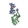 4rixS S: Starting model for refinement |
|---|---|
| Similar structure data |


-Links
 PDBj
PDBj


- Assembly
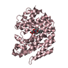
| Deposited unit | 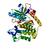
| ||||||||
|---|---|---|---|---|---|---|---|---|---|
| 1 |
| ||||||||
| Unit cell |
|
-Components
| #1: Protein | Mass: 37949.613 Da / Num. of mol.: 1 / Fragment: kinase domain (UNP residues 866-1191) Source method: isolated from a genetically manipulated source Source: (gene. exp.) Caenorhabditis elegans (invertebrata) / Gene: let-23, kin-7, ZK1067.1 / Plasmid: pFASTbac HTa / Production host:   Spodoptera frugiperda (fall armyworm) / Strain (production host): Sf9 Spodoptera frugiperda (fall armyworm) / Strain (production host): Sf9References: UniProt: P24348, receptor protein-tyrosine kinase |
|---|---|
| #2: Chemical | ChemComp-ANP /   Mass: 506.196 Da / Num. of mol.: 1 / Source method: obtained synthetically / Formula: C10H17N6O12P3 / Comment: AMP-PNP, energy-carrying molecule analogue*YM Mass: 506.196 Da / Num. of mol.: 1 / Source method: obtained synthetically / Formula: C10H17N6O12P3 / Comment: AMP-PNP, energy-carrying molecule analogue*YM |
| #3: Chemical | ChemComp-MG /   Mass: 24.305 Da / Num. of mol.: 1 / Source method: obtained synthetically / Formula: Mg Mass: 24.305 Da / Num. of mol.: 1 / Source method: obtained synthetically / Formula: Mg |
| #4: Water | ChemComp-HOH / Water Mass: 18.015 Da / Num. of mol.: 11 / Source method: isolated from a natural source / Formula: H2O Mass: 18.015 Da / Num. of mol.: 11 / Source method: isolated from a natural source / Formula: H2O |
-Experimental details
-Experiment
| Experiment | Method: X-RAY DIFFRACTION / Number of used crystals: 1 |
|---|
- Sample preparation
| Crystal | Density Matthews: 2.62 Å3/Da / Density % sol: 53.13 % |
|---|---|
| Crystal grow | Temperature: 293 K / Method: vapor diffusion, hanging drop / pH: 7.5 Details: 20% PEG5000 MME, 0.20 M sodium chloride, 0.10 M HEPES, pH 7.5 |
-Data collection
| Diffraction | Mean temperature: 100 K |
|---|---|
| Diffraction source | Source: SYNCHROTRON / Site: ALS / Beamline: 8.3.1 / Wavelength: 1.11587 Å |
| Detector | Type: ADSC QUANTUM 315r / Detector: CCD / Date: Apr 30, 2015 |
| Radiation | Monochromator: double crystal Si(111) / Protocol: SINGLE WAVELENGTH / Monochromatic (M) / Laue (L): M / Scattering type: x-ray |
| Radiation wavelength | Wavelength: 1.11587 Å / Relative weight: 1 |
| Reflection | Resolution: 2.386→63 Å / Num. obs: 16320 / % possible obs: 98.8 % / Redundancy: 6.4 % / Biso Wilson estimate: 65.15 Å2 / Rmerge(I) obs: 0.1 / Net I/σ(I): 10.8 |
| Reflection shell | Resolution: 2.386→2.46 Å / Rmerge(I) obs: 0.89 / Mean I/σ(I) obs: 1.1 / % possible all: 98.5 |
- Processing
| Software |
| |||||||||||||||||||||||||||||||||||||||||||||||||
|---|---|---|---|---|---|---|---|---|---|---|---|---|---|---|---|---|---|---|---|---|---|---|---|---|---|---|---|---|---|---|---|---|---|---|---|---|---|---|---|---|---|---|---|---|---|---|---|---|---|---|
| Refinement | Method to determine structure: MOLECULAR REPLACEMENT Starting model: PDB entry 4RIX Resolution: 2.386→40.936 Å / SU ML: 0.29 / Cross valid method: FREE R-VALUE / σ(F): 1.34 / Phase error: 31.31 / Stereochemistry target values: ML
| |||||||||||||||||||||||||||||||||||||||||||||||||
| Solvent computation | Shrinkage radii: 0.9 Å / VDW probe radii: 1.11 Å / Solvent model: FLAT BULK SOLVENT MODEL | |||||||||||||||||||||||||||||||||||||||||||||||||
| Refinement step | Cycle: LAST / Resolution: 2.386→40.936 Å
| |||||||||||||||||||||||||||||||||||||||||||||||||
| Refine LS restraints |
| |||||||||||||||||||||||||||||||||||||||||||||||||
| LS refinement shell |
| |||||||||||||||||||||||||||||||||||||||||||||||||
| Refinement TLS params. | Method: refined / Origin x: -27.0873 Å / Origin y: 4.4576 Å / Origin z: -19.5396 Å
| |||||||||||||||||||||||||||||||||||||||||||||||||
| Refinement TLS group | Selection details: all |
