 Movie
Movie Controller


+ Open data

- Basic information
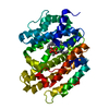
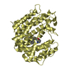
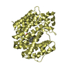
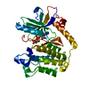
| Entry | Database: PDB / ID: 1ezf | ||||||
|---|---|---|---|---|---|---|---|
| Title | CRYSTAL STRUCTURE OF HUMAN SQUALENE SYNTHASE | ||||||
 Components Components | FARNESYL-DIPHOSPHATE FARNESYLTRANSFERASE | ||||||
 Keywords Keywords | TRANSFERASE / isoprenoid synthase fold / all alpha-helix | ||||||
| Function / homology |  Function and homology information Function and homology informationfarnesyl diphosphate metabolic process / squalene synthase / farnesyl-diphosphate farnesyltransferase activity / squalene synthase activity / Cholesterol biosynthesis / steroid biosynthetic process / cholesterol biosynthetic process / Activation of gene expression by SREBF (SREBP) / PPARA activates gene expression / endoplasmic reticulum membrane ...farnesyl diphosphate metabolic process / squalene synthase / farnesyl-diphosphate farnesyltransferase activity / squalene synthase activity / Cholesterol biosynthesis / steroid biosynthetic process / cholesterol biosynthetic process / Activation of gene expression by SREBF (SREBP) / PPARA activates gene expression / endoplasmic reticulum membrane / endoplasmic reticulum / membrane / metal ion bindingSimilarity search - Function | ||||||
| Biological species |  Homo sapiens (human) Homo sapiens (human) | ||||||
| Method | X-RAY DIFFRACTION / MIRAS / Resolution: 2.15 Å | ||||||
 Authors Authors | Pandit, J. / Danley, D.E. / Schulte, G.K. / Mazzalupo, S.M. / Pauly, T.A. / Hayward, C.M. / Hamanaka, E.S. / Thompson, J.F. / Harwood, H.J. | ||||||
 Citation Citation | Journal: J.Biol.Chem. / Year: 2000 Title: Crystal structure of human squalene synthase. A key enzyme in cholesterol biosynthesis. Authors: Pandit, J. / Danley, D.E. / Schulte, G.K. / Mazzalupo, S. / Pauly, T.A. / Hayward, C.M. / Hamanaka, E.S. / Thompson, J.F. / Harwood Jr., H.J. | ||||||
| History |
|
- Structure visualization
| Structure viewer | Molecule: MolmilJmol/JSmol |
|---|
- Downloads & links
-Download
| PDBx/mmCIF format | 1ezf.cif.gz | 210.6 KB | Display | PDBx/mmCIF format |
|---|---|---|---|---|
| PDB format | pdb1ezf.ent.gz | 170.3 KB | Display | PDB format |
| PDBx/mmJSON format | 1ezf.json.gz | Tree view | PDBx/mmJSON format | |
| Others |  Other downloads Other downloads |
-Validation report
| Arichive directory | https://data.pdbj.org/pub/pdb/validation_reports/ez/1ezfftp://data.pdbj.org/pub/pdb/validation_reports/ez/1ezf | HTTPS FTP |
|---|
-Related structure data
| Similar structure data |
|---|


-Links
 PDBj
PDBj

- Assembly
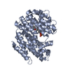
| Deposited unit | 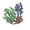
| ||||||||
|---|---|---|---|---|---|---|---|---|---|
| 1 | 
| ||||||||
| 2 | 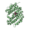
| ||||||||
| 3 | 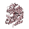
| ||||||||
| Unit cell |
| ||||||||
| Details | The biological assembly is a monomer. |
-Components
| #1: Protein | / SQUALENE SYNTHETASE Mass: 39221.805 Da / Num. of mol.: 3 / Fragment: RESIDUES 31-370 / Mutation: Q33R, D34N, S38N Source method: isolated from a genetically manipulated source Source: (gene. exp.) Homo sapiens (human) / Plasmid: PHSS9 / Production host:  Escherichia coli (E. coli) / References: UniProt: P37268, squalene synthase Escherichia coli (E. coli) / References: UniProt: P37268, squalene synthase#2: Chemical |   Mass: 567.029 Da / Num. of mol.: 3 / Source method: obtained synthetically / Formula: C30H31ClN2O7 Mass: 567.029 Da / Num. of mol.: 3 / Source method: obtained synthetically / Formula: C30H31ClN2O7#3: Water | ChemComp-HOH / | Water Mass: 18.015 Da / Num. of mol.: 410 / Source method: isolated from a natural source / Formula: H2O Mass: 18.015 Da / Num. of mol.: 410 / Source method: isolated from a natural source / Formula: H2O |
|---|
-Experimental details
-Experiment
| Experiment | Method: X-RAY DIFFRACTION / Number of used crystals: 1 |
|---|
- Sample preparation
| Crystal | Density Matthews: 2.32 Å3/Da / Density % sol: 47 % | ||||||||||||||||||||||||||||||||||||||||||
|---|---|---|---|---|---|---|---|---|---|---|---|---|---|---|---|---|---|---|---|---|---|---|---|---|---|---|---|---|---|---|---|---|---|---|---|---|---|---|---|---|---|---|---|
| Crystal grow | Temperature: 293 K / Method: vapor diffusion, hanging drop / pH: 5.6 Details: 25-30% PEG 4000, 0.1M sodium citrate, pH5.6, 0.2M ammonium acetate , VAPOR DIFFUSION, HANGING DROP, temperature 20K | ||||||||||||||||||||||||||||||||||||||||||
| Crystal grow | *PLUS | ||||||||||||||||||||||||||||||||||||||||||
| Components of the solutions | *PLUS
|
-Data collection
| Diffraction | Mean temperature: 100 K |
|---|---|
| Diffraction source | Source: ROTATING ANODE / Type: RIGAKU RU200 / Wavelength: 1.5418 |
| Detector | Type: RIGAKU RAXIS IIC / Detector: IMAGE PLATE / Date: Nov 1, 1995 |
| Radiation | Monochromator: Graphite / Protocol: SINGLE WAVELENGTH / Monochromatic (M) / Laue (L): M / Scattering type: x-ray |
| Radiation wavelength | Wavelength: 1.5418 Å / Relative weight: 1 |
| Reflection | Resolution: 2.15→40 Å / Observed criterion σ(I): -3 / Biso Wilson estimate: 26 Å2 / Rmerge(I) obs: 0.042 / Net I/σ(I): 35 |
| Reflection shell | Resolution: 2.15→2.23 Å / Rmerge(I) obs: 0.141 / Mean I/σ(I) obs: 7.2 / % possible all: 84.9 |
| Reflection | *PLUS Num. obs: 55224 / % possible obs: 96 % |
| Reflection shell | *PLUS % possible obs: 84.9 % |
- Processing
| Software |
| |||||||||||||||||||||||||
|---|---|---|---|---|---|---|---|---|---|---|---|---|---|---|---|---|---|---|---|---|---|---|---|---|---|---|
| Refinement | Method to determine structure: MIRAS / Resolution: 2.15→40 Å / Data cutoff high absF: 1000000 / Data cutoff low absF: 0.1 / Cross valid method: Free-R / σ(F): 2 / Stereochemistry target values: Engh & Huber
| |||||||||||||||||||||||||
| Refinement step | Cycle: LAST / Resolution: 2.15→40 Å
| |||||||||||||||||||||||||
| Refine LS restraints |
| |||||||||||||||||||||||||
| LS refinement shell | Resolution: 2.15→2.23 Å / Total num. of bins used: 10
|
