[English] 日本語
Yorodumi
- PDB-4ino: The crystal structure of Helicobacter pylori Ceue (HP1561) -

+
Open data


ID or keywords:

Loading...

-
Basic information

Entry
Database: PDB / ID: 4ino
TitleThe crystal structure of Helicobacter pylori Ceue (HP1561)
ComponentsNickel (II) ABC transporter, periplasmic nickel-binding protein
Keywords
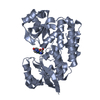
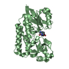
TRANSPORT PROTEIN / heme-binding protein / Fe transport / ABC transporter substrate binding protein / periplasmic space
Function / homologyABC transporter periplasmic binding domain / Periplasmic binding protein / Iron siderophore/cobalamin periplasmic-binding domain profile. / Nitrogenase molybdenum iron protein domain / cellular response to iron ion / Rossmann fold / 3-Layer(aba) Sandwich / Alpha Beta / Iron (III) ABC transporter, periplasmic iron-bindin gprotein
Function and homology information
Biological speciesHelicobacter pylori (bacteria)
MethodX-RAY DIFFRACTION / SYNCHROTRON / SAD / Resolution: 1.65 Å
AuthorsShaik, M.M. / Cendron, L. / Zanotti, G.
CitationJournal: Mol.Microbiol. / Year: 2014
Title: Helicobacter pylori periplasmic receptor CeuE (HP1561) modulates its nickel affinity via organic metallophores.
Authors: Shaik, M.M. / Cendron, L. / Salamina, M. / Ruzzene, M. / Zanotti, G.
History
DepositionJan 5, 2013Deposition site: RCSB / Processing site: RCSB
Revision 1.0Jan 8, 2014Provider: repository / Type: Initial release
Revision 1.1Jan 15, 2014Group: Database references
Revision 1.2Feb 26, 2014Group: Database references
Revision 1.3Jan 31, 2018Group: Experimental preparation / Category: exptl_crystal_grow / Item: _exptl_crystal_grow.temp
Revision 1.4Jul 17, 2019Group: Data collection / Refinement description / Category: software
Item: _software.classification / _software.name / _software.version
Revision 1.5Feb 28, 2024Group: Data collection / Database references / Refinement description
Category: chem_comp_atom / chem_comp_bond ...chem_comp_atom / chem_comp_bond / database_2 / struct_ncs_dom_lim / struct_ref_seq_dif
Item: _database_2.pdbx_DOI / _database_2.pdbx_database_accession ..._database_2.pdbx_DOI / _database_2.pdbx_database_accession / _struct_ncs_dom_lim.beg_auth_comp_id / _struct_ncs_dom_lim.beg_label_asym_id / _struct_ncs_dom_lim.beg_label_comp_id / _struct_ncs_dom_lim.beg_label_seq_id / _struct_ncs_dom_lim.end_auth_comp_id / _struct_ncs_dom_lim.end_label_asym_id / _struct_ncs_dom_lim.end_label_comp_id / _struct_ncs_dom_lim.end_label_seq_id / _struct_ref_seq_dif.details

-
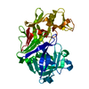
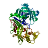
Structure visualization

Structure viewerMolecule:
MolmilJmol/JSmol

Downloads & links

-
Assembly

Deposited unit
A: Nickel (II) ABC transporter, periplasmic nickel-binding protein
B: Nickel (II) ABC transporter, periplasmic nickel-binding protein


Theoretical massNumber of molelcules
Total (without water)75,0592
Polymers75,0592
Non-polymers00
Water8,881493
1
A: Nickel (II) ABC transporter, periplasmic nickel-binding protein


Theoretical massNumber of molelcules
Total (without water)37,5291
Polymers37,5291
Non-polymers00
Water181
TypeNameSymmetry operationNumber
identity operation1_555x,y,z1
MethodPISA
2
B: Nickel (II) ABC transporter, periplasmic nickel-binding protein


Theoretical massNumber of molelcules
Total (without water)37,5291
Polymers37,5291
Non-polymers00
Water181
TypeNameSymmetry operationNumber
identity operation1_555x,y,z1
MethodPISA
Unit cell
Length a, b, c (Å)67.370, 87.020, 105.900
Angle α, β, γ (deg.)90.00, 90.00, 90.00
Int Tables number19
Space group name H-MP212121
Noncrystallographic symmetry (NCS)NCS domain:
IDEns-IDDetails
11A
21B

NCS domain segments:

Component-ID: 1 / Ens-ID: 1 / Beg auth comp-ID: GLN / Beg label comp-ID: GLN / End auth comp-ID: HIS / End label comp-ID: HIS / Refine code: 5 / Auth seq-ID: 34 - 335 / Label seq-ID: 1 - 302

Dom-IDAuth asym-IDLabel asym-ID
1AA
2BB

-
Components

#1: Protein Nickel (II) ABC transporter, periplasmic nickel-binding protein / CEUE


Mass: 37529.477 Da / Num. of mol.: 2 / Fragment: UNP residues 32-333 / Mutation: V102A
Source method: isolated from a genetically manipulated source
Source: (gene. exp.) Helicobacter pylori (bacteria) / Strain: G27 / Gene: HPG27_1499 / Plasmid: pET101-HP1561 / Production host: Escherichia coli (E. coli) / Strain (production host): BL21(DE3) / References: UniProt: B5Z9J2
#2: Water ChemComp-HOH / water / Water


Mass: 18.015 Da / Num. of mol.: 493 / Source method: isolated from a natural source / Formula: H2O

-
Experimental details

-
Experiment

ExperimentMethod: X-RAY DIFFRACTION / Number of used crystals: 1

-
Sample preparation

CrystalDensity Matthews: 2.07 Å3/Da / Density % sol: 40.52 %
Crystal growTemperature: 293 K / pH: 8
Details: 0.1 M SPG buffer, 25% w/v PEG1500, pH 8.0, VAPOR DIFFUSION, temperature 393K

-
Data collection

Diffraction
IDMean temperature (K)Crystal-ID
11001
21001
Diffraction source
SourceSiteBeamlineIDWavelength
SYNCHROTRONESRF BM1410.97627
SYNCHROTRONESRF ID2920.9792
Detector
TypeIDDetectorDate
MARMOSAIC 225 mm CCD1CCDAug 31, 2011
PSI PILATUS 6M2PIXELMay 5, 2011
Radiation
IDMonochromatorProtocolMonochromatic (M) / Laue (L)Scattering typeWavelength-ID
1Si(111)SINGLE WAVELENGTHMx-ray1
2Si(111)SINGLE WAVELENGTHMx-ray1
Radiation wavelength
IDWavelength (Å)Relative weight
10.976271
20.97921
ReflectionResolution: 1.65→47.59 Å / Num. obs: 73090 / % possible obs: 97 % / Observed criterion σ(I): 0 / Redundancy: 4.1 % / Rmerge(I) obs: 0.054 / Net I/σ(I): 10.2
Reflection shellResolution: 1.65→1.74 Å / Redundancy: 4.2 % / Rmerge(I) obs: 0.51 / Mean I/σ(I) obs: 2.3 / % possible all: 96.8

-
Processing

Software
NameVersionClassification
PHENIX1.8.2_1309refinement
REFMAC5.5.0109refinement
MOSFLMdata reduction
SCALAdata scaling
AutoSolphasing
RefinementMethod to determine structure: SAD / Resolution: 1.65→45.234 Å / Cor.coef. Fo:Fc: 0.961 / Cor.coef. Fo:Fc free: 0.946 / SU ML: 0.18 / σ(F): 1.33 / Phase error: 22.46 / Stereochemistry target values: ML / Details: HYDROGENS HAVE BEEN ADDED IN THE BINDING POSITIONS
RfactorNum. reflection% reflection
Rfree0.2246 3661 5.01 %
Rwork0.1932 --
obs0.1948 73002 96.65 %
Solvent computationShrinkage radii: 0.9 Å / VDW probe radii: 1.11 Å / Solvent model: FLAT BULK SOLVENT MODEL
Displacement parametersBiso mean: 29.34 Å2
Baniso -1Baniso -2Baniso -3
1-0.01 Å2-0 Å20 Å2
2---0.01 Å2-0 Å2
3---0 Å2
Refinement stepCycle: LAST / Resolution: 1.65→45.234 Å
ProteinNucleic acidLigandSolventTotal
Num. atoms4796 0 0 493 5289
Refine LS restraints
Refine-IDTypeDev idealNumber
X-RAY DIFFRACTIONf_bond_d0.0074904
X-RAY DIFFRACTIONf_angle_d1.1496620
X-RAY DIFFRACTIONf_dihedral_angle_d13.7541840
X-RAY DIFFRACTIONf_chiral_restr0.082746
X-RAY DIFFRACTIONf_plane_restr0.005832
Refine LS restraints NCS

Dom-ID: 1 / Auth asym-ID: A / Ens-ID: 1 / Refine-ID: X-RAY DIFFRACTION

NumberTypeRms dev position (Å)Weight position
1208medium positional0.20.5
1190loose positional0.445
1208medium thermal2.352
1190loose thermal2.2710
LS refinement shellResolution: 1.65→1.69 Å / Total num. of bins used: 20
RfactorNum. reflection% reflection
Rfree0.31 263 -
Rwork0.289 5064 -
obs--96.4 %

+
About Yorodumi

-
News

-
Feb 9, 2022. New format data for meta-information of EMDB entries

New format data for meta-information of EMDB entries

  • Version 3 of the EMDB header file is now the official format.
  • The previous official version 1.9 will be removed from the archive.

Related info.:EMDB header

External links:wwPDB to switch to version 3 of the EMDB data model

-
Aug 12, 2020. Covid-19 info

Covid-19 info

URL: https://pdbj.org/emnavi/covid19.php

New page: Covid-19 featured information page in EM Navigator.

Related info.:Covid-19 info / Mar 5, 2020. Novel coronavirus structure data

+
Mar 5, 2020. Novel coronavirus structure data

Novel coronavirus structure data

Related info.:Yorodumi Speices / Aug 12, 2020. Covid-19 info

External links:COVID-19 featured content - PDBj / Molecule of the Month (242):Coronavirus Proteases

+
Jan 31, 2019. EMDB accession codes are about to change! (news from PDBe EMDB page)

EMDB accession codes are about to change! (news from PDBe EMDB page)

  • The allocation of 4 digits for EMDB accession codes will soon come to an end. Whilst these codes will remain in use, new EMDB accession codes will include an additional digit and will expand incrementally as the available range of codes is exhausted. The current 4-digit format prefixed with “EMD-” (i.e. EMD-XXXX) will advance to a 5-digit format (i.e. EMD-XXXXX), and so on. It is currently estimated that the 4-digit codes will be depleted around Spring 2019, at which point the 5-digit format will come into force.
  • The EM Navigator/Yorodumi systems omit the EMD- prefix.

Related info.:Q: What is EMD? / ID/Accession-code notation in Yorodumi/EM Navigator

External links:EMDB Accession Codes are Changing Soon! / Contact to PDBj

+
Jul 12, 2017. Major update of PDB

Major update of PDB

  • wwPDB released updated PDB data conforming to the new PDBx/mmCIF dictionary.
  • This is a major update changing the version number from 4 to 5, and with Remediation, in which all the entries are updated.
  • In this update, many items about electron microscopy experimental information are reorganized (e.g. em_software).
  • Now, EM Navigator and Yorodumi are based on the updated data.

External links:wwPDB Remediation / Enriched Model Files Conforming to OneDep Data Standards Now Available in the PDB FTP Archive

-
Yorodumi

Thousand views of thousand structures

  • Yorodumi is a browser for structure data from EMDB, PDB, SASBDB, etc.
  • This page is also the successor to EM Navigator detail page, and also detail information page/front-end page for Omokage search.
  • The word "yorodu" (or yorozu) is an old Japanese word meaning "ten thousand". "mi" (miru) is to see.

Related info.:EMDB / PDB / SASBDB / Comparison of 3 databanks / Yorodumi Search / Aug 31, 2016. New EM Navigator & Yorodumi / Yorodumi Papers / Jmol/JSmol / Function and homology information / Changes in new EM Navigator and Yorodumi

Read more