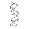
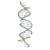
 Movie
Movie Controller


+ Open data

- Basic information
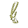
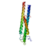
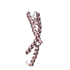
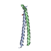
| Entry | Database: PDB / ID: 5vy7 | ||||||
|---|---|---|---|---|---|---|---|
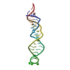
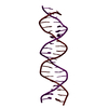
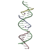
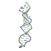
| Title | A self-assembling L-form DNA crystal lattice | ||||||
 Components Components |
| ||||||
 Keywords Keywords |  DNA / DNA nanotechnology / self-assembly / DNA structure DNA / DNA nanotechnology / self-assembly / DNA structure | ||||||
| Function / homology | DNA / DNA (> 10) Function and homology information Function and homology information | ||||||
| Biological species | synthetic construct (others) | ||||||
| Method | X-RAY DIFFRACTION / SYNCHROTRON / SAD / Resolution: 3 Å | ||||||
 Authors Authors | Simmons, C.R. / Yan, H. | ||||||
 Citation Citation | Journal: J. Am. Chem. Soc. / Year: 2017 Title: Tuning the Cavity Size and Chirality of Self-Assembling 3D DNA Crystals. Authors: Simmons, C.R. / Zhang, F. / MacCulloch, T. / Fahmi, N. / Stephanopoulos, N. / Liu, Y. / Seeman, N.C. / Yan, H. | ||||||
| History |
|
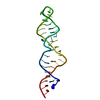
- Structure visualization
| Structure viewer | Molecule: MolmilJmol/JSmol |
|---|
- Downloads & links
-Download
| PDBx/mmCIF format | 5vy7.cif.gz | 31.3 KB | Display | PDBx/mmCIF format |
|---|---|---|---|---|
| PDB format | pdb5vy7.ent.gz | 27.9 KB | Display | PDB format |
| PDBx/mmJSON format | 5vy7.json.gz | Tree view | PDBx/mmJSON format | |
| Others |  Other downloads Other downloads |
-Validation report
| Arichive directory | https://data.pdbj.org/pub/pdb/validation_reports/vy/5vy7ftp://data.pdbj.org/pub/pdb/validation_reports/vy/5vy7 | HTTPS FTP |
|---|
-Related structure data


-Links
 PDBj
PDBj


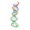
- Assembly
| Deposited unit | 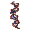
| ||||||||
|---|---|---|---|---|---|---|---|---|---|
| 1 |
| ||||||||
| Unit cell |
|
-Components
| #1: DNA chain | Mass: 6466.201 Da / Num. of mol.: 1 / Source method: obtained synthetically / Source: (synth.) synthetic construct (others) |
|---|---|
| #2: DNA chain | Mass: 1769.193 Da / Num. of mol.: 1 / Source method: obtained synthetically / Source: (synth.) synthetic construct (others) |
| #3: DNA chain | Mass: 2432.614 Da / Num. of mol.: 1 / Source method: obtained synthetically / Source: (synth.) synthetic construct (others) |
| #4: DNA chain | Mass: 2129.409 Da / Num. of mol.: 1 / Source method: obtained synthetically / Source: (synth.) synthetic construct (others) |
-Experimental details
-Experiment
| Experiment | Method: X-RAY DIFFRACTION / Number of used crystals: 1 |
|---|
- Sample preparation
| Crystal | Density Matthews: 5.91 Å3/Da / Density % sol: 79.2 % |
|---|---|
| Crystal grow | Temperature: 298 K / Method: vapor diffusion, sitting drop Details: 50 mM cacodylate pH 6.5, 50 mM MgCl2, 2.0 mM cobalt(III)hexamine, and 1.5 M lithium sulfate |
-Data collection
| Diffraction | Mean temperature: 100 K |
|---|---|
| Diffraction source | Source: SYNCHROTRON / Site: ALS  / Beamline: 8.2.2 / Wavelength: 1 Å / Beamline: 8.2.2 / Wavelength: 1 Å |
| Detector | Type: ADSC QUANTUM 315r / Detector: CCD / Date: Jan 13, 2016 |
| Radiation | Protocol: SINGLE WAVELENGTH / Monochromatic (M) / Laue (L): M / Scattering type: x-ray |
| Radiation wavelength | Wavelength: 1 Å / Relative weight: 1 |
| Reflection | Resolution: 3→34.292 Å / Num. obs: 4949 / % possible obs: 86.1 % / Redundancy: 10.7 % / Rmerge(I) obs: 0.11 / Net I/σ(I): 23.9 |
| Reflection shell | Resolution: 3→3.05 Å / Redundancy: 9.5 % / Rmerge(I) obs: 0.467 / Mean I/σ(I) obs: 2.6 / % possible all: 53.7 |
- Processing
| Software |
| ||||||||||||||||||||||||
|---|---|---|---|---|---|---|---|---|---|---|---|---|---|---|---|---|---|---|---|---|---|---|---|---|---|
| Refinement | Method to determine structure: SAD / Resolution: 3→34.29 Å / SU ML: 0.25 / Cross valid method: FREE R-VALUE / σ(F): 2.01 / Phase error: 37.7
| ||||||||||||||||||||||||
| Solvent computation | Shrinkage radii: 0.9 Å / VDW probe radii: 1.11 Å | ||||||||||||||||||||||||
| Refinement step | Cycle: LAST / Resolution: 3→34.29 Å
| ||||||||||||||||||||||||
| Refine LS restraints |
| ||||||||||||||||||||||||
| LS refinement shell |
|
