 Movie
Movie Controller


+ Open data

- Basic information
| Entry | Database: PDB / ID: 6akm | ||||||
|---|---|---|---|---|---|---|---|
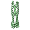
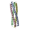
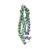
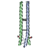
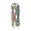
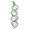
| Title | Crystal structure of SLMAP-SIKE1 complex | ||||||
 Components Components |
| ||||||
 Keywords Keywords |  PROTEIN BINDING / Coiled-coil domain / heterotetramer PROTEIN BINDING / Coiled-coil domain / heterotetramer | ||||||
| Function / homology |  Function and homology information Function and homology informationregulation of membrane depolarization during cardiac muscle cell action potential / regulation of voltage-gated sodium channel activity / regulation of sodium ion transmembrane transport / TRAF6 mediated IRF7 activation / smooth endoplasmic reticulum / muscle contraction / protein localization to plasma membrane / sarcolemma / small GTPase binding / SARS-CoV-1 activates/modulates innate immune responses ...regulation of membrane depolarization during cardiac muscle cell action potential / regulation of voltage-gated sodium channel activity / regulation of sodium ion transmembrane transport / TRAF6 mediated IRF7 activation / smooth endoplasmic reticulum / muscle contraction / protein localization to plasma membrane / sarcolemma / small GTPase binding / SARS-CoV-1 activates/modulates innate immune responses / TRAF3-dependent IRF activation pathway / centrosome / protein kinase binding / SARS-CoV-2 activates/modulates innate and adaptive immune responses / plasma membrane / cytosolSimilarity search - Function | ||||||
| Biological species |  Homo sapiens (human) Homo sapiens (human) | ||||||
| Method | X-RAY DIFFRACTION / SYNCHROTRON / MOLECULAR REPLACEMENT / Resolution: 2.3 Å | ||||||
 Authors Authors | Ma, J. / Chen, M. / Zhou, Z.C. | ||||||
 Citation Citation | Journal: Cell Discov / Year: 2019 Title: Architecture, substructures, and dynamic assembly of STRIPAK complexes in Hippo signaling. Authors: Tang, Y. / Chen, M. / Zhou, L. / Ma, J. / Li, Y. / Zhang, H. / Shi, Z. / Xu, Q. / Zhang, X. / Gao, Z. / Zhao, Y. / Cheng, Y. / Jiao, S. / Zhou, Z. | ||||||
| History |
|
- Structure visualization
| Structure viewer | Molecule: MolmilJmol/JSmol |
|---|
- Downloads & links
-Download
| PDBx/mmCIF format | 6akm.cif.gz | 54.1 KB | Display | PDBx/mmCIF format |
|---|---|---|---|---|
| PDB format | pdb6akm.ent.gz | 37.6 KB | Display | PDB format |
| PDBx/mmJSON format | 6akm.json.gz | Tree view | PDBx/mmJSON format | |
| Others |  Other downloads Other downloads |
-Validation report
| Arichive directory | https://data.pdbj.org/pub/pdb/validation_reports/ak/6akmftp://data.pdbj.org/pub/pdb/validation_reports/ak/6akm | HTTPS FTP |
|---|
-Related structure data
| Related structure data |  6akkSC 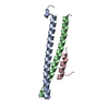 6aklC S: Starting model for refinement C: citing same article ( |
|---|---|
| Similar structure data |


-Links
 PDBj
PDBj


- Assembly
| Deposited unit | 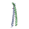
| ||||||||
|---|---|---|---|---|---|---|---|---|---|
| 1 | 
| ||||||||
| Unit cell |
| ||||||||
| Components on special symmetry positions |
|
-Components
| #1: Protein | Mass: 7184.042 Da / Num. of mol.: 1 Source method: isolated from a genetically manipulated source Source: (gene. exp.) Homo sapiens (human) / Gene: SIKE1, SIKE / Production host:  Escherichia coli BL21(DE3) (bacteria) / Strain (production host): BL21 (DE3) / Variant (production host): CodonPlus / References: UniProt: Q9BRV8 Escherichia coli BL21(DE3) (bacteria) / Strain (production host): BL21 (DE3) / Variant (production host): CodonPlus / References: UniProt: Q9BRV8 |
|---|---|
| #2: Protein | Mass: 7421.357 Da / Num. of mol.: 1 Source method: isolated from a genetically manipulated source Source: (gene. exp.) Homo sapiens (human) / Gene: SLMAP, KIAA1601, SLAP, UNQ1847/PRO3577 / Production host: Escherichia coli BL21(DE3) (bacteria) / Strain (production host): BL21 (DE3) / Variant (production host): CodonPlus / References: UniProt: Q14BN4 |
| #3: Chemical | ChemComp-GOL / Glycerol  Mass: 92.094 Da / Num. of mol.: 1 / Source method: obtained synthetically / Formula: C3H8O3 Mass: 92.094 Da / Num. of mol.: 1 / Source method: obtained synthetically / Formula: C3H8O3 |
| #4: Water | ChemComp-HOH / Water Mass: 18.015 Da / Num. of mol.: 35 / Source method: isolated from a natural source / Formula: H2O Mass: 18.015 Da / Num. of mol.: 35 / Source method: isolated from a natural source / Formula: H2O |
-Experimental details
-Experiment
| Experiment | Method: X-RAY DIFFRACTION / Number of used crystals: 1 |
|---|
- Sample preparation
| Crystal | Density Matthews: 2.7 Å3/Da / Density % sol: 54.49 % |
|---|---|
| Crystal grow | Temperature: 289 K / Method: vapor diffusion, sitting drop / Details: 0.1 M sodium citrate, pH 5.5; 15% (w/v) PEG 6000 |
-Data collection
| Diffraction | Mean temperature: 100 K / Serial crystal experiment: N |
|---|---|
| Diffraction source | Source: SYNCHROTRON / Site: SSRF  / Beamline: BL18U1 / Wavelength: 0.97853 Å / Beamline: BL18U1 / Wavelength: 0.97853 Å |
| Detector | Type: DECTRIS PILATUS3 6M / Detector: PIXEL / Date: Dec 11, 2017 |
| Radiation | Protocol: SINGLE WAVELENGTH / Monochromatic (M) / Laue (L): M / Scattering type: x-ray |
| Radiation wavelength | Wavelength: 0.97853 Å / Relative weight: 1 |
| Reflection | Resolution: 2.3→50 Å / Num. obs: 7336 / % possible obs: 100 % / Redundancy: 18.8 % / Rmerge(I) obs: 0.094 / Rpim(I) all: 0.022 / Rrim(I) all: 0.097 / Net I/σ(I): 21.6 |
| Reflection shell | Resolution: 2.3→2.38 Å / Redundancy: 19 % / Rmerge(I) obs: 0.821 / Mean I/σ(I) obs: 5.2 / Num. unique obs: 713 / CC1/2: 0.948 / Rpim(I) all: 0.193 / Rrim(I) all: 0.844 / % possible all: 100 |
- Processing
| Software |
| |||||||||||||||||||||||||||||||||||||||||||||||||||||||||||||||||||||||||||
|---|---|---|---|---|---|---|---|---|---|---|---|---|---|---|---|---|---|---|---|---|---|---|---|---|---|---|---|---|---|---|---|---|---|---|---|---|---|---|---|---|---|---|---|---|---|---|---|---|---|---|---|---|---|---|---|---|---|---|---|---|---|---|---|---|---|---|---|---|---|---|---|---|---|---|---|---|
| Refinement | Method to determine structure: MOLECULAR REPLACEMENT Starting model: 6AKK Resolution: 2.3→42.08 Å / SU ML: 0.25 / Cross valid method: FREE R-VALUE / σ(F): 1.34 / Phase error: 25.09
| |||||||||||||||||||||||||||||||||||||||||||||||||||||||||||||||||||||||||||
| Solvent computation | Shrinkage radii: 0.9 Å / VDW probe radii: 1.11 Å | |||||||||||||||||||||||||||||||||||||||||||||||||||||||||||||||||||||||||||
| Refinement step | Cycle: LAST / Resolution: 2.3→42.08 Å
| |||||||||||||||||||||||||||||||||||||||||||||||||||||||||||||||||||||||||||
| Refine LS restraints |
| |||||||||||||||||||||||||||||||||||||||||||||||||||||||||||||||||||||||||||
| LS refinement shell |
| |||||||||||||||||||||||||||||||||||||||||||||||||||||||||||||||||||||||||||
| Refinement TLS params. | Method: refined / Refine-ID: X-RAY DIFFRACTION
| |||||||||||||||||||||||||||||||||||||||||||||||||||||||||||||||||||||||||||
| Refinement TLS group |
|
