 Movie
Movie Controller

[English] 日本語
 Yorodumi
- PDB-5ou9: Crystal structure of Glycoprotein VI in complex with collagen-pep... -
+ Open data

- Basic information
| Entry | Database: PDB / ID: 5ou9 | ||||||
|---|---|---|---|---|---|---|---|
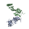
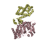
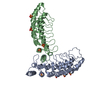
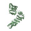
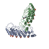
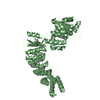
| Title | Crystal structure of Glycoprotein VI in complex with collagen-peptide (GPO)3 | ||||||
 Components Components |
| ||||||
 Keywords Keywords |  BLOOD CLOTTING / Platelet / glycoprotein / GPVI / collagen-binding / platelet activation / GPO3 / CRP BLOOD CLOTTING / Platelet / glycoprotein / GPVI / collagen-binding / platelet activation / GPO3 / CRP | ||||||
| Function / homology |  Function and homology information Function and homology informationcellular response to fluoride / collagen type I trimer / tooth mineralization / cellular response to vitamin E / collagen type IV trimer / Anchoring fibril formation / Crosslinking of collagen fibrils / collagen biosynthetic process / Collagen chain trimerization / Enhanced cleavage of VWF variant by ADAMTS13 ...cellular response to fluoride / collagen type I trimer / tooth mineralization / cellular response to vitamin E / collagen type IV trimer / Anchoring fibril formation / Crosslinking of collagen fibrils / collagen biosynthetic process / Collagen chain trimerization / Enhanced cleavage of VWF variant by ADAMTS13 / Defective VWF cleavage by ADAMTS13 variant / Defective VWF binding to collagen type I / platelet-derived growth factor binding / bone trabecula formation / extracellular matrix structural constituent conferring tensile strength / Enhanced binding of GP1BA variant to VWF multimer:collagen / Defective binding of VWF variant to GPIb:IX:V / intramembranous ossification / Extracellular matrix organization / embryonic skeletal system development / Collagen biosynthesis and modifying enzymes / cartilage development involved in endochondral bone morphogenesis / tetraspanin-enriched microdomain / skin morphogenesis / collagen-activated tyrosine kinase receptor signaling pathway / collagen-activated signaling pathway / Platelet Adhesion to exposed collagen / endochondral ossification / positive regulation of platelet aggregation / cellular response to fibroblast growth factor stimulus / collagen fibril organization / negative regulation of cell-substrate adhesion / response to steroid hormone / face morphogenesis / Scavenging by Class A Receptors / skin development / MET activates PTK2 signaling / Assembly of collagen fibrils and other multimeric structures / Syndecan interactions / GP1b-IX-V activation signalling / enzyme-linked receptor protein signaling pathway / blood vessel development / RUNX2 regulates osteoblast differentiation / Platelet Aggregation (Plug Formation) / Collagen degradation / protein localization to nucleus / Non-integrin membrane-ECM interactions / ECM proteoglycans / response to hyperoxia / Integrin cell surface interactions / positive regulation of epithelial to mesenchymal transition / response to mechanical stimulus / cellular response to retinoic acid / GPVI-mediated activation cascade / cellular response to epidermal growth factor stimulus / response to cAMP / cellular response to transforming growth factor beta stimulus / collagen binding / visual perception / extracellular matrix organization / ossification / protein tyrosine kinase binding / secretory granule / skeletal system development / Cell surface interactions at the vascular wall / cellular response to glucose stimulus / sensory perception of sound / cellular response to amino acid stimulus / response to insulin / response to hydrogen peroxide / platelet activation / osteoblast differentiation / cellular response to mechanical stimulus / transmembrane signaling receptor activity / Immunoregulatory interactions between a Lymphoid and a non-Lymphoid cell / positive regulation of canonical Wnt signaling pathway / protein transport / response to estradiol / signaling receptor activity / cellular response to tumor necrosis factor / collagen-containing extracellular matrix / protease binding / positive regulation of cell migration / response to xenobiotic stimulus / membrane raft / endoplasmic reticulum lumen / positive regulation of DNA-templated transcription / cell surface / extracellular space / extracellular exosome / extracellular region / identical protein binding / metal ion binding / plasma membrane / cytoplasmSimilarity search - Function | ||||||
| Biological species |  Homo sapiens (human) Homo sapiens (human) | ||||||
| Method | X-RAY DIFFRACTION / SYNCHROTRON / MOLECULAR REPLACEMENT / Resolution: 2.5 Å | ||||||
 Authors Authors | Feitsma, L.J. / Huizinga, E.G. | ||||||
| Funding support |  Netherlands, 1items Netherlands, 1items
| ||||||
 Citation Citation | Journal: To be Published Title: Structural insights into collagen-binding by platelet receptor Glycoprotein VI Authors: Feitsma, L.J. / Brondijk, T.H.C. / Jarvis, G. / Hagemans, D. / Bihan, D. / Jerah, N. / Versteeg, M. / Farndale, R.W. / Huizinga, E.G. | ||||||
| History |
|
- Structure visualization
| Structure viewer | Molecule: MolmilJmol/JSmol |
|---|
- Downloads & links
-Download
| PDBx/mmCIF format | 5ou9.cif.gz | 173.9 KB | Display | PDBx/mmCIF format |
|---|---|---|---|---|
| PDB format | pdb5ou9.ent.gz | 139.9 KB | Display | PDB format |
| PDBx/mmJSON format | 5ou9.json.gz | Tree view | PDBx/mmJSON format | |
| Others |  Other downloads Other downloads |
-Validation report
| Arichive directory | https://data.pdbj.org/pub/pdb/validation_reports/ou/5ou9ftp://data.pdbj.org/pub/pdb/validation_reports/ou/5ou9 | HTTPS FTP |
|---|
-Related structure data
| Related structure data |  5ou7SC S: Starting model for refinement C: citing same article ( |
|---|---|
| Similar structure data |


-Links
 PDBj
PDBj


- Assembly
| Deposited unit | 
| ||||||||
|---|---|---|---|---|---|---|---|---|---|
| 1 |
| ||||||||
| Unit cell |
|
-Components
-Protein / Protein/peptide / Sugars , 3 types, 7 molecules ABCDE
| #1: Protein | Mass: 19719.215 Da / Num. of mol.: 2 / Mutation: -102-105 -131-136 Source method: isolated from a genetically manipulated source Source: (gene. exp.) Homo sapiens (human) / Gene: GP6 / Cell line (production host): HEK293-EBNA1-S / Production host: Homo sapiens (human) / References: UniProt: Q9HCN6#2: Protein/peptide | Mass: 1824.985 Da / Num. of mol.: 3 / Source method: obtained synthetically / Source: (synth.) Homo sapiens (human) / References: UniProt: P02452*PLUS#3: Sugar | N-Acetylglucosamine Type: D-saccharide, beta linking / Mass: 221.208 Da / Num. of mol.: 2 Type: D-saccharide, beta linking / Mass: 221.208 Da / Num. of mol.: 2Source method: isolated from a genetically manipulated source Formula: C8H15NO6 |
|---|
-Non-polymers , 3 types, 44 molecules 


| #4: Chemical | Polyethylene glycol Mass: 194.226 Da / Num. of mol.: 2 / Source method: obtained synthetically / Formula: C8H18O5 / Comment: precipitant*YM Mass: 194.226 Da / Num. of mol.: 2 / Source method: obtained synthetically / Formula: C8H18O5 / Comment: precipitant*YM#5: Chemical | ChemComp-CL / | Chloride Mass: 35.453 Da / Num. of mol.: 1 / Source method: obtained synthetically / Formula: Cl Mass: 35.453 Da / Num. of mol.: 1 / Source method: obtained synthetically / Formula: Cl#6: Water | ChemComp-HOH / | WaterMass: 18.015 Da / Num. of mol.: 41 / Source method: isolated from a natural source / Formula: H2O |
|---|
-Experimental details
-Experiment
| Experiment | Method: X-RAY DIFFRACTION / Number of used crystals: 1 |
|---|
- Sample preparation
| Crystal | Density Matthews: 3.1 Å3/Da / Density % sol: 60.33 % |
|---|---|
| Crystal grow | Temperature: 293 K / Method: vapor diffusion, sitting drop / pH: 9 / Details: 0.1 M MMT-buffer pH 9.0 25% (w/v) PEG1500 |
-Data collection
| Diffraction | Mean temperature: 100 K |
|---|---|
| Diffraction source | Source: SYNCHROTRON / Site: ESRF  / Beamline: ID23-1 / Wavelength: 0.97242 Å / Beamline: ID23-1 / Wavelength: 0.97242 Å |
| Detector | Type: ADSC QUANTUM 315r / Detector: CCD / Date: Oct 8, 2013 |
| Radiation | Protocol: SINGLE WAVELENGTH / Monochromatic (M) / Laue (L): M / Scattering type: x-ray |
| Radiation wavelength | Wavelength: 0.97242 Å / Relative weight: 1 |
| Reflection | Resolution: 2.5→79.93 Å / Num. obs: 21273 / % possible obs: 99.9 % / Redundancy: 6.4 % / Biso Wilson estimate: 56.4 Å2 / CC1/2: 0.984 / Rmerge(I) obs: 0.113 / Net I/σ(I): 8.5 |
| Reflection shell | Resolution: 2.5→2.61 Å / Redundancy: 6.8 % / Rmerge(I) obs: 0.971 / Num. unique obs: 2503 / CC1/2: 0.646 / % possible all: 100 |
- Processing
| Software |
| ||||||||||||||||||||||||||||||||||||||||||||||||||||||||||||||||||||||||||||||||||||||||||||||||||||||||||||||||||||||||||||||||||||||||||||||||||||||||||||||||||||||||||||||||||||||
|---|---|---|---|---|---|---|---|---|---|---|---|---|---|---|---|---|---|---|---|---|---|---|---|---|---|---|---|---|---|---|---|---|---|---|---|---|---|---|---|---|---|---|---|---|---|---|---|---|---|---|---|---|---|---|---|---|---|---|---|---|---|---|---|---|---|---|---|---|---|---|---|---|---|---|---|---|---|---|---|---|---|---|---|---|---|---|---|---|---|---|---|---|---|---|---|---|---|---|---|---|---|---|---|---|---|---|---|---|---|---|---|---|---|---|---|---|---|---|---|---|---|---|---|---|---|---|---|---|---|---|---|---|---|---|---|---|---|---|---|---|---|---|---|---|---|---|---|---|---|---|---|---|---|---|---|---|---|---|---|---|---|---|---|---|---|---|---|---|---|---|---|---|---|---|---|---|---|---|---|---|---|---|---|
| Refinement | Method to determine structure: MOLECULAR REPLACEMENT Starting model: 5OU7 Resolution: 2.5→79.93 Å / Cor.coef. Fo:Fc: 0.94 / Cor.coef. Fo:Fc free: 0.92 / SU B: 24.728 / SU ML: 0.251 / Cross valid method: THROUGHOUT / ESU R: 0.384 / ESU R Free: 0.264 / Details: HYDROGENS HAVE BEEN ADDED IN THE RIDING POSITIONS
| ||||||||||||||||||||||||||||||||||||||||||||||||||||||||||||||||||||||||||||||||||||||||||||||||||||||||||||||||||||||||||||||||||||||||||||||||||||||||||||||||||||||||||||||||||||||
| Solvent computation | Ion probe radii: 0.8 Å / Shrinkage radii: 0.8 Å / VDW probe radii: 1.2 Å | ||||||||||||||||||||||||||||||||||||||||||||||||||||||||||||||||||||||||||||||||||||||||||||||||||||||||||||||||||||||||||||||||||||||||||||||||||||||||||||||||||||||||||||||||||||||
| Displacement parameters | Biso mean: 69.524 Å2
| ||||||||||||||||||||||||||||||||||||||||||||||||||||||||||||||||||||||||||||||||||||||||||||||||||||||||||||||||||||||||||||||||||||||||||||||||||||||||||||||||||||||||||||||||||||||
| Refinement step | Cycle: 1 / Resolution: 2.5→79.93 Å
| ||||||||||||||||||||||||||||||||||||||||||||||||||||||||||||||||||||||||||||||||||||||||||||||||||||||||||||||||||||||||||||||||||||||||||||||||||||||||||||||||||||||||||||||||||||||
| Refine LS restraints |
|