 Movie
Movie Controller

[English] 日本語
 Yorodumi
- PDB-5ou7: Crystal structure of the Glycoprotein VI loop truncation mutant P... -
+ Open data

- Basic information
| Entry | Database: PDB / ID: 5ou7 | ||||||
|---|---|---|---|---|---|---|---|
| Title | Crystal structure of the Glycoprotein VI loop truncation mutant PAVS-PAPYKN | ||||||
 Components Components | Platelet glycoprotein VI | ||||||
 Keywords Keywords |  BLOOD CLOTTING / Platelet / glycoprotein / GPVI / collagen-binding / platelet activation BLOOD CLOTTING / Platelet / glycoprotein / GPVI / collagen-binding / platelet activation | ||||||
| Function / homology |  Function and homology information Function and homology informationtetraspanin-enriched microdomain / collagen-activated signaling pathway / Platelet Adhesion to exposed collagen / positive regulation of platelet aggregation / enzyme-linked receptor protein signaling pathway / GPVI-mediated activation cascade / collagen binding / protein tyrosine kinase binding / Cell surface interactions at the vascular wall / platelet activation ...tetraspanin-enriched microdomain / collagen-activated signaling pathway / Platelet Adhesion to exposed collagen / positive regulation of platelet aggregation / enzyme-linked receptor protein signaling pathway / GPVI-mediated activation cascade / collagen binding / protein tyrosine kinase binding / Cell surface interactions at the vascular wall / platelet activation / transmembrane signaling receptor activity / signaling receptor activity / membrane raft / cell surface / extracellular exosome / plasma membraneSimilarity search - Function | ||||||
| Biological species |  Homo sapiens (human) Homo sapiens (human) | ||||||
| Method | X-RAY DIFFRACTION / SYNCHROTRON / MOLECULAR REPLACEMENT / Resolution: 1.9 Å | ||||||
 Authors Authors | Feitsma, L.J. / Huizinga, E.G. | ||||||
| Funding support |  Netherlands, 1items Netherlands, 1items
| ||||||
 Citation Citation | Journal: To Be Published Title: Structural insights into collagen-binding by platelet receptor Glycoprotein VI Authors: Feitsma, L.J. / Brondijk, T.H.C. / Jarvis, G. / Hagemans, D. / Bihan, D. / Jerah, N. / Versteeg, M. / Farndale, R.W. / Huizinga, E.G. | ||||||
| History |
|
- Structure visualization
| Structure viewer | Molecule: MolmilJmol/JSmol |
|---|
- Downloads & links
-Download
| PDBx/mmCIF format | 5ou7.cif.gz | 295.6 KB | Display | PDBx/mmCIF format |
|---|---|---|---|---|
| PDB format | pdb5ou7.ent.gz | 241.3 KB | Display | PDB format |
| PDBx/mmJSON format | 5ou7.json.gz | Tree view | PDBx/mmJSON format | |
| Others |  Other downloads Other downloads |
-Validation report
| Arichive directory | https://data.pdbj.org/pub/pdb/validation_reports/ou/5ou7ftp://data.pdbj.org/pub/pdb/validation_reports/ou/5ou7 | HTTPS FTP |
|---|
-Related structure data
| Related structure data |  5ou9C  2gi7S S: Starting model for refinement C: citing same article ( |
|---|---|
| Similar structure data |


-Links
 PDBj
PDBj


- Assembly
| Deposited unit | 
| ||||||||||||||||||||||||||||||||||||||||||||||||||||||||||||||||||||||||||||||
|---|---|---|---|---|---|---|---|---|---|---|---|---|---|---|---|---|---|---|---|---|---|---|---|---|---|---|---|---|---|---|---|---|---|---|---|---|---|---|---|---|---|---|---|---|---|---|---|---|---|---|---|---|---|---|---|---|---|---|---|---|---|---|---|---|---|---|---|---|---|---|---|---|---|---|---|---|---|---|---|
| 1 | 
| ||||||||||||||||||||||||||||||||||||||||||||||||||||||||||||||||||||||||||||||
| 2 | 
| ||||||||||||||||||||||||||||||||||||||||||||||||||||||||||||||||||||||||||||||
| 3 | 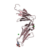
| ||||||||||||||||||||||||||||||||||||||||||||||||||||||||||||||||||||||||||||||
| 4 | 
| ||||||||||||||||||||||||||||||||||||||||||||||||||||||||||||||||||||||||||||||
| Unit cell |
| ||||||||||||||||||||||||||||||||||||||||||||||||||||||||||||||||||||||||||||||
| Noncrystallographic symmetry (NCS) | NCS domain:
NCS domain segments: Component-ID: 1 / Beg auth comp-ID: SER / Beg label comp-ID: SER / Refine code: 2
NCS ensembles :
NCS oper:
|
-Components
-Protein / Sugars , 2 types, 8 molecules ABCD
| #1: Protein | Mass: 19719.215 Da / Num. of mol.: 4 / Mutation: -102-105 -131-136 Source method: isolated from a genetically manipulated source Source: (gene. exp.) Homo sapiens (human) / Gene: GP6 / Cell line (production host): HEK293-EBNA1-S / Production host: Homo sapiens (human) / References: UniProt: Q9HCN6#5: Sugar | ChemComp-NAG / N-Acetylglucosamine Type: D-saccharide, beta linking / Mass: 221.208 Da / Num. of mol.: 4 Type: D-saccharide, beta linking / Mass: 221.208 Da / Num. of mol.: 4Source method: isolated from a genetically manipulated source Formula: C8H15NO6 |
|---|
-Non-polymers , 4 types, 362 molecules 


| #2: Chemical | ChemComp-CL / Chloride Mass: 35.453 Da / Num. of mol.: 14 / Source method: obtained synthetically / Formula: Cl Mass: 35.453 Da / Num. of mol.: 14 / Source method: obtained synthetically / Formula: Cl#3: Chemical | Phosphate Mass: 94.971 Da / Num. of mol.: 2 / Source method: obtained synthetically / Formula: PO4 Mass: 94.971 Da / Num. of mol.: 2 / Source method: obtained synthetically / Formula: PO4#4: Chemical | Polyethylene glycol Mass: 266.331 Da / Num. of mol.: 2 / Source method: obtained synthetically / Formula: C12H26O6 Mass: 266.331 Da / Num. of mol.: 2 / Source method: obtained synthetically / Formula: C12H26O6#6: Water | ChemComp-HOH / | WaterMass: 18.015 Da / Num. of mol.: 344 / Source method: isolated from a natural source / Formula: H2O |
|---|
-Experimental details
-Experiment
| Experiment | Method: X-RAY DIFFRACTION / Number of used crystals: 1 |
|---|
- Sample preparation
| Crystal | Density Matthews: 2.5 Å3/Da / Density % sol: 50.87 % |
|---|---|
| Crystal grow | Temperature: 293 K / Method: vapor diffusion, sitting drop / pH: 4 Details: 0.1 M phosphate-citrate buffer pH 4.0 40% (v/v) PEG-300 |
-Data collection
| Diffraction | Mean temperature: 100 K |
|---|---|
| Diffraction source | Source: SYNCHROTRON / Site: SLS  / Beamline: X06SA / Wavelength: 0.99999 Å / Beamline: X06SA / Wavelength: 0.99999 Å |
| Detector | Type: DECTRIS EIGER X 16M / Detector: PIXEL / Date: Aug 22, 2013 |
| Radiation | Protocol: SINGLE WAVELENGTH / Monochromatic (M) / Laue (L): M / Scattering type: x-ray |
| Radiation wavelength | Wavelength: 0.99999 Å / Relative weight: 1 |
| Reflection | Resolution: 1.9→113.81 Å / Num. obs: 58873 / % possible obs: 95.1 % / Redundancy: 2.1 % / Biso Wilson estimate: 25.6 Å2 / CC1/2: 0.99 / Rmerge(I) obs: 0.081 / Net I/σ(I): 5 |
| Reflection shell | Resolution: 1.9→1.94 Å / Redundancy: 2.1 % / Rmerge(I) obs: 1.08 / Mean I/σ(I) obs: 1.1 / Num. unique obs: 3656 / CC1/2: 0.62 / % possible all: 93.3 |
- Processing
| Software |
| ||||||||||||||||||||||||||||||||||||||||||||||||||||||||||||||||||||||||||||||||||||||||||||||||||||||||||||||||||||||||||||||||||||||||||||||||||||||||||||||||||||||||||||||||||||||
|---|---|---|---|---|---|---|---|---|---|---|---|---|---|---|---|---|---|---|---|---|---|---|---|---|---|---|---|---|---|---|---|---|---|---|---|---|---|---|---|---|---|---|---|---|---|---|---|---|---|---|---|---|---|---|---|---|---|---|---|---|---|---|---|---|---|---|---|---|---|---|---|---|---|---|---|---|---|---|---|---|---|---|---|---|---|---|---|---|---|---|---|---|---|---|---|---|---|---|---|---|---|---|---|---|---|---|---|---|---|---|---|---|---|---|---|---|---|---|---|---|---|---|---|---|---|---|---|---|---|---|---|---|---|---|---|---|---|---|---|---|---|---|---|---|---|---|---|---|---|---|---|---|---|---|---|---|---|---|---|---|---|---|---|---|---|---|---|---|---|---|---|---|---|---|---|---|---|---|---|---|---|---|---|
| Refinement | Method to determine structure: MOLECULAR REPLACEMENT Starting model: 2GI7 Resolution: 1.9→113.81 Å / Cor.coef. Fo:Fc: 0.959 / Cor.coef. Fo:Fc free: 0.939 / SU B: 10.313 / SU ML: 0.143 / Cross valid method: THROUGHOUT / ESU R: 0.192 / ESU R Free: 0.169 / Details: HYDROGENS HAVE BEEN ADDED IN THE RIDING POSITIONS
| ||||||||||||||||||||||||||||||||||||||||||||||||||||||||||||||||||||||||||||||||||||||||||||||||||||||||||||||||||||||||||||||||||||||||||||||||||||||||||||||||||||||||||||||||||||||
| Solvent computation | Ion probe radii: 0.8 Å / Shrinkage radii: 0.8 Å / VDW probe radii: 1.2 Å | ||||||||||||||||||||||||||||||||||||||||||||||||||||||||||||||||||||||||||||||||||||||||||||||||||||||||||||||||||||||||||||||||||||||||||||||||||||||||||||||||||||||||||||||||||||||
| Displacement parameters | Biso mean: 33.189 Å2
| ||||||||||||||||||||||||||||||||||||||||||||||||||||||||||||||||||||||||||||||||||||||||||||||||||||||||||||||||||||||||||||||||||||||||||||||||||||||||||||||||||||||||||||||||||||||
| Refinement step | Cycle: 1 / Resolution: 1.9→113.81 Å
| ||||||||||||||||||||||||||||||||||||||||||||||||||||||||||||||||||||||||||||||||||||||||||||||||||||||||||||||||||||||||||||||||||||||||||||||||||||||||||||||||||||||||||||||||||||||
| Refine LS restraints |
|