 Movie
Movie Controller


+ Open data

- Basic information
| Entry | Database: PDB / ID: 3b7b | ||||||
|---|---|---|---|---|---|---|---|
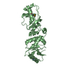
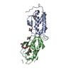
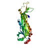
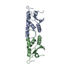
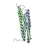
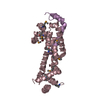
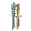
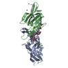
| Title | EuHMT1 (Glp) Ankyrin Repeat Domain (Structure 1) | ||||||
 Components Components | Euchromatic histone-lysine N-methyltransferase 1 | ||||||
 Keywords Keywords |  TRANSFERASE / Ankyrin repeat / Alternative splicing / ANK repeat / Chromatin regulator / Methyltransferase / Nucleus / Phosphorylation / Polymorphism / S-adenosyl-L-methionine TRANSFERASE / Ankyrin repeat / Alternative splicing / ANK repeat / Chromatin regulator / Methyltransferase / Nucleus / Phosphorylation / Polymorphism / S-adenosyl-L-methionine | ||||||
| Function / homology |  Function and homology information Function and homology information[histone H3]-lysine9 N-methyltransferase / histone H3K27 methyltransferase activity / peptidyl-lysine monomethylation / histone H3K9 methyltransferase activity / histone H3K9me2 methyltransferase activity / peptidyl-lysine dimethylation / protein-lysine N-methyltransferase activity / DNA methylation-dependent heterochromatin formation / C2H2 zinc finger domain binding / Transcriptional Regulation by E2F6 ...[histone H3]-lysine9 N-methyltransferase / histone H3K27 methyltransferase activity / peptidyl-lysine monomethylation / histone H3K9 methyltransferase activity / histone H3K9me2 methyltransferase activity / peptidyl-lysine dimethylation / protein-lysine N-methyltransferase activity / DNA methylation-dependent heterochromatin formation / C2H2 zinc finger domain binding / Transcriptional Regulation by E2F6 / regulation of embryonic development / Transcriptional Regulation by VENTX / response to fungicide / Transferases; Transferring one-carbon groups; Methyltransferases / transcription corepressor binding / methyltransferase activity / Regulation of TP53 Activity through Methylation / PKMTs methylate histone lysines / p53 binding / positive regulation of cold-induced thermogenesis / chromatin organization / Senescence-Associated Secretory Phenotype (SASP) / nuclear body / negative regulation of DNA-templated transcription / chromatin / negative regulation of transcription by RNA polymerase II / zinc ion binding / nucleoplasm / nucleusSimilarity search - Function | ||||||
| Biological species |  Homo sapiens (human) Homo sapiens (human) | ||||||
| Method | X-RAY DIFFRACTION / SYNCHROTRON / MOLECULAR REPLACEMENT / Resolution: 2.99 Å | ||||||
 Authors Authors | Collins, R.E. / Horton, J.R. / Cheng, X. | ||||||
 Citation Citation | Journal: Nat.Struct.Mol.Biol. / Year: 2008 Title: The ankyrin repeats of G9a and GLP histone methyltransferases are mono- and dimethyllysine binding modules Authors: Collins, R.E. / Northrop, J.P. / Horton, J.R. / Lee, D.Y. / Zhang, X. / Stallcup, M.R. / Cheng, X. | ||||||
| History |
|
- Structure visualization
| Structure viewer | Molecule: MolmilJmol/JSmol |
|---|
- Downloads & links
-Download
| PDBx/mmCIF format | 3b7b.cif.gz | 97.6 KB | Display | PDBx/mmCIF format |
|---|---|---|---|---|
| PDB format | pdb3b7b.ent.gz | 75.5 KB | Display | PDB format |
| PDBx/mmJSON format | 3b7b.json.gz | Tree view | PDBx/mmJSON format | |
| Others |  Other downloads Other downloads |
-Validation report
| Arichive directory | https://data.pdbj.org/pub/pdb/validation_reports/b7/3b7bftp://data.pdbj.org/pub/pdb/validation_reports/b7/3b7b | HTTPS FTP |
|---|
-Related structure data


-Links
 PDBj
PDBj


- Assembly
| Deposited unit | 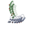
| ||||||||
|---|---|---|---|---|---|---|---|---|---|
| 1 | 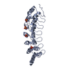
| ||||||||
| 2 | 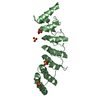
| ||||||||
| Unit cell |
|
-Components
| #1: Protein | Mass: 26175.650 Da / Num. of mol.: 2 / Fragment: Ankyrin repeat domains 2-7 Source method: isolated from a genetically manipulated source Source: (gene. exp.) Homo sapiens (human) / Gene: EHMT1, EUHMTASE1, KIAA1876 / Production host:  Escherichia coli (E. coli) / Strain (production host): BL21 (DE3) CodonPlus Escherichia coli (E. coli) / Strain (production host): BL21 (DE3) CodonPlusReferences: UniProt: Q9H9B1, histone-lysine N-methyltransferase#2: Chemical | ChemComp-SO4 / Sulfate  Mass: 96.063 Da / Num. of mol.: 8 / Source method: obtained synthetically / Formula: SO4 Mass: 96.063 Da / Num. of mol.: 8 / Source method: obtained synthetically / Formula: SO4#3: Water | ChemComp-HOH / | Water Mass: 18.015 Da / Num. of mol.: 28 / Source method: isolated from a natural source / Formula: H2O Mass: 18.015 Da / Num. of mol.: 28 / Source method: isolated from a natural source / Formula: H2O |
|---|
-Experimental details
-Experiment
| Experiment | Method: X-RAY DIFFRACTION / Number of used crystals: 1 |
|---|
- Sample preparation
| Crystal | Density Matthews: 3.51 Å3/Da / Density % sol: 64.93 % |
|---|---|
| Crystal grow | Temperature: 289 K / Method: vapor diffusion, sitting drop / pH: 8.5 Details: 1.5-2.0 M Lithium Sulfate, 1-4% Peg 400 or 550, 0.1 M Tris buffer pH 8.5, VAPOR DIFFUSION, SITTING DROP, temperature 289K |
-Data collection
| Diffraction | Mean temperature: 100 K |
|---|---|
| Diffraction source | Source: SYNCHROTRON / Site: APS  / Beamline: 22-ID / Wavelength: 0.97625 Å / Beamline: 22-ID / Wavelength: 0.97625 Å |
| Detector | Type: MARMOSAIC 300 mm CCD / Detector: CCD / Date: Apr 12, 2007 |
| Radiation | Monochromator: Si (220) / Protocol: SINGLE WAVELENGTH / Monochromatic (M) / Laue (L): M / Scattering type: x-ray |
| Radiation wavelength | Wavelength: 0.97625 Å / Relative weight: 1 |
| Reflection | Resolution: 2.99→28.75 Å / Num. all: 16000 / Num. obs: 15965 / % possible obs: 99.8 % / Observed criterion σ(F): -3 / Observed criterion σ(I): -3 / Redundancy: 11.9 % / Biso Wilson estimate: 32.5 Å2 / Rmerge(I) obs: 0.123 / Net I/σ(I): 13.7 |
| Reflection shell | Resolution: 2.99→3.1 Å / Redundancy: 11 % / Rmerge(I) obs: 0.535 / Num. unique all: 1564 / % possible all: 99.7 |
- Processing
| Software |
| |||||||||||||||||||||||||
|---|---|---|---|---|---|---|---|---|---|---|---|---|---|---|---|---|---|---|---|---|---|---|---|---|---|---|
| Refinement | Method to determine structure: MOLECULAR REPLACEMENT Starting model: Generated Model from Phyre server Resolution: 2.99→28.75 Å / Isotropic thermal model: ISOTROPIC / Cross valid method: THROUGOUT / σ(F): 0 / σ(I): 0 / Stereochemistry target values: Engh & Huber
| |||||||||||||||||||||||||
| Displacement parameters | Biso mean: 47.4 Å2
| |||||||||||||||||||||||||
| Refine analyze |
| |||||||||||||||||||||||||
| Refinement step | Cycle: LAST / Resolution: 2.99→28.75 Å
| |||||||||||||||||||||||||
| Refine LS restraints |
| |||||||||||||||||||||||||
| LS refinement shell | Resolution: 2.99→3.1 Å / Rfactor Rfree error: 0.049
|
