 Movie
Movie Controller

[English] 日本語
 Yorodumi
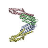
- PDB-2b3t: Structure of complex between E. coli translation termination fact... -
+ Open data

- Basic information
| Entry | Database: PDB / ID: 2b3t | ||||||
|---|---|---|---|---|---|---|---|
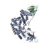
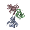
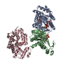
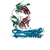
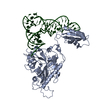
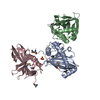
| Title | Structure of complex between E. coli translation termination factor RF1 and the PrmC methyltransferase | ||||||
 Components Components |
| ||||||
 Keywords Keywords |  TRANSLATION / release factor / translation termination / methylation / conformational changes TRANSLATION / release factor / translation termination / methylation / conformational changes | ||||||
| Function / homology |  Function and homology information Function and homology informationpeptide chain release factor N5-glutamine methyltransferase / protein-(glutamine-N5) methyltransferase activity / protein-glutamine N-methyltransferase activity / peptidyl-glutamine methylation / protein methyltransferase activity / translation release factor activity, codon specific / protein methylation / S-adenosylmethionine-dependent methyltransferase activity / translational termination / ribosome binding ...peptide chain release factor N5-glutamine methyltransferase / protein-(glutamine-N5) methyltransferase activity / protein-glutamine N-methyltransferase activity / peptidyl-glutamine methylation / protein methyltransferase activity / translation release factor activity, codon specific / protein methylation / S-adenosylmethionine-dependent methyltransferase activity / translational termination / ribosome binding / regulation of gene expression / nucleic acid binding / cytosolSimilarity search - Function | ||||||
| Biological species |  Escherichia coli (E. coli) Escherichia coli (E. coli) | ||||||
| Method | X-RAY DIFFRACTION / SYNCHROTRON / MOLECULAR REPLACEMENT / Resolution: 3.1 Å | ||||||
 Authors Authors | Graille, M. / Heurgue-Hamard, V. / Champ, S. / Mora, L. / Scrima, N. / Ulryck, N. / van Tilbeurgh, H. / Buckingham, R.H. | ||||||
 Citation Citation | Journal: Mol.Cell / Year: 2005 Title: Molecular basis for bacterial class I release factor methylation by PrmC Authors: Graille, M. / Heurgue-Hamard, V. / Champ, S. / Mora, L. / Scrima, N. / Ulryck, N. / van Tilbeurgh, H. / Buckingham, R.H. | ||||||
| History |
|
- Structure visualization
| Structure viewer | Molecule: MolmilJmol/JSmol |
|---|
- Downloads & links
-Download
| PDBx/mmCIF format | 2b3t.cif.gz | 126.3 KB | Display | PDBx/mmCIF format |
|---|---|---|---|---|
| PDB format | pdb2b3t.ent.gz | 97.6 KB | Display | PDB format |
| PDBx/mmJSON format | 2b3t.json.gz | Tree view | PDBx/mmJSON format | |
| Others |  Other downloads Other downloads |
-Validation report
| Arichive directory | https://data.pdbj.org/pub/pdb/validation_reports/b3/2b3tftp://data.pdbj.org/pub/pdb/validation_reports/b3/2b3t | HTTPS FTP |
|---|
-Related structure data
| Related structure data | |
|---|---|
| Similar structure data |


-Links
 PDBj
PDBj
- Assembly
| Deposited unit | 
| ||||||||
|---|---|---|---|---|---|---|---|---|---|
| 1 |
| ||||||||
| Unit cell |
| ||||||||
| Details | The heterodimer present in the asymetric unit corresponds to the biological unit. |
-Components
| #1: Protein | Mass: 30877.789 Da / Num. of mol.: 1 Source method: isolated from a genetically manipulated source Source: (gene. exp.) Escherichia coli (E. coli) / Gene: hemK / Production host: Escherichia coli (E. coli)References: UniProt: P37186, UniProt: P0ACC1*PLUS, Transferases; Transferring one-carbon groups; Methyltransferases |
|---|---|
| #2: Protein | Mass: 40559.281 Da / Num. of mol.: 1 Source method: isolated from a genetically manipulated source Source: (gene. exp.) Escherichia coli (E. coli) / Gene: prfA, sueB, uar / Production host: Escherichia coli (E. coli) / References: UniProt: P0A7I0 |
| #3: Chemical | ChemComp-SAH / S-Adenosyl-L-homocysteine  Type: L-peptide linking / Mass: 384.411 Da / Num. of mol.: 1 / Source method: obtained synthetically / Formula: C14H20N6O5S Type: L-peptide linking / Mass: 384.411 Da / Num. of mol.: 1 / Source method: obtained synthetically / Formula: C14H20N6O5S |
-Experimental details
-Experiment
| Experiment | Method: X-RAY DIFFRACTION / Number of used crystals: 1 |
|---|
- Sample preparation
| Crystal | Density Matthews: 2.7 Å3/Da / Density % sol: 55 % |
|---|---|
| Crystal grow | Temperature: 291 K / Method: vapor diffusion, sitting drop / pH: 7.2 Details: ammonium sulfate, NaCl, glycerol, Na Hepes, pH 7.2, VAPOR DIFFUSION, SITTING DROP, temperature 291K |
-Data collection
| Diffraction | Mean temperature: 100 K | ||||||||||||||||||||||||||||||||||||||||||||||||||||||||||||
|---|---|---|---|---|---|---|---|---|---|---|---|---|---|---|---|---|---|---|---|---|---|---|---|---|---|---|---|---|---|---|---|---|---|---|---|---|---|---|---|---|---|---|---|---|---|---|---|---|---|---|---|---|---|---|---|---|---|---|---|---|---|
| Diffraction source | Source: SYNCHROTRON / Site: ESRF  / Beamline: ID23-1 / Wavelength: 0.97 Å / Beamline: ID23-1 / Wavelength: 0.97 Å | ||||||||||||||||||||||||||||||||||||||||||||||||||||||||||||
| Detector | Type: MARRESEARCH / Detector: CCD / Date: Jul 3, 2004 | ||||||||||||||||||||||||||||||||||||||||||||||||||||||||||||
| Radiation | Monochromator: Si 111 CHANNEL / Protocol: SINGLE WAVELENGTH / Monochromatic (M) / Laue (L): M / Scattering type: x-ray | ||||||||||||||||||||||||||||||||||||||||||||||||||||||||||||
| Radiation wavelength | Wavelength: 0.97 Å / Relative weight: 1 | ||||||||||||||||||||||||||||||||||||||||||||||||||||||||||||
| Reflection | Av σ(I) over netI: 12.25 / Number: 52659 / Rmerge(I) obs: 10.4 / D res high: 3.1 Å / Num. obs: 14361 / % possible obs: 96.3 | ||||||||||||||||||||||||||||||||||||||||||||||||||||||||||||
| Diffraction reflection shell |
| ||||||||||||||||||||||||||||||||||||||||||||||||||||||||||||
| Reflection | Resolution: 3.1→20 Å / Num. obs: 14670 / % possible obs: 96.3 % / Rmerge(I) obs: 0.104 / Net I/σ(I): 12.25 | ||||||||||||||||||||||||||||||||||||||||||||||||||||||||||||
| Reflection shell | Resolution: 3.1→3.2 Å / % possible obs: 91.8 % / Rmerge(I) obs: 0.474 / Mean I/σ(I) obs: 3.82 / Num. measured obs: 4594 / Num. unique obs: 1229 |
- Processing
| Software |
| ||||||||||||||||||||||||||||||||||||
|---|---|---|---|---|---|---|---|---|---|---|---|---|---|---|---|---|---|---|---|---|---|---|---|---|---|---|---|---|---|---|---|---|---|---|---|---|---|
| Refinement | Method to determine structure: MOLECULAR REPLACEMENT / Resolution: 3.1→20 Å / σ(F): 0 / Stereochemistry target values: Engh & Huber
| ||||||||||||||||||||||||||||||||||||
| Solvent computation | Bsol: 29.444 Å2 | ||||||||||||||||||||||||||||||||||||
| Displacement parameters | Biso mean: 55.406 Å2
| ||||||||||||||||||||||||||||||||||||
| Refinement step | Cycle: LAST / Resolution: 3.1→20 Å
| ||||||||||||||||||||||||||||||||||||
| Refine LS restraints |
| ||||||||||||||||||||||||||||||||||||
| Xplor file |
|
