 Movie
Movie Controller

[English] 日本語
 Yorodumi
- PDB-3tup: Crystal structure of human mitochondrial PheRS complexed with tRN... -
+ Open data

- Basic information
| Entry | Database: PDB / ID: 3tup | ||||||
|---|---|---|---|---|---|---|---|
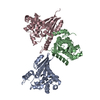
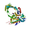
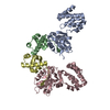
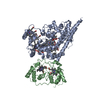
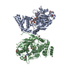
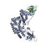
| Title | Crystal structure of human mitochondrial PheRS complexed with tRNAPhe in the active open state | ||||||
 Components Components |
| ||||||
 Keywords Keywords | LIGASE/RNA / class II aaRS / RRM fold /  aminoacylation / mitochondria / LIGASE-RNA complex aminoacylation / mitochondria / LIGASE-RNA complex | ||||||
| Function / homology |  Function and homology informationphenylalanine-tRNA ligase / Mitochondrial tRNA aminoacylation / phenylalanine-tRNA ligase activity / phenylalanyl-tRNA aminoacylation / tRNA aminoacylation for protein translation / tRNA processing / tRNA binding / mitochondrial matrix / mitochondrion / ATP binding / cytoplasm Function and homology informationphenylalanine-tRNA ligase / Mitochondrial tRNA aminoacylation / phenylalanine-tRNA ligase activity / phenylalanyl-tRNA aminoacylation / tRNA aminoacylation for protein translation / tRNA processing / tRNA binding / mitochondrial matrix / mitochondrion / ATP binding / cytoplasmSimilarity search - Function | ||||||
| Biological species |  Homo sapiens (human) Homo sapiens (human)  Thermus thermophilus (bacteria) Thermus thermophilus (bacteria) | ||||||
| Method | X-RAY DIFFRACTION / SYNCHROTRON / MOLECULAR REPLACEMENT / Resolution: 3.05 Å | ||||||
 Authors Authors | Safro, M. / Klipcan, L. / Moor, N. / Finarov, I. / Kessler, N. / Sukhanova, M. | ||||||
 Citation Citation | Journal: J.Mol.Biol. / Year: 2012 Title: Crystal Structure of Human Mitochondrial PheRS Complexed with tRNA(Phe) in the Active "Open" State. Authors: Klipcan, L. / Moor, N. / Finarov, I. / Kessler, N. / Sukhanova, M. / Safro, M.G. | ||||||
| History |
|
- Structure visualization
| Structure viewer | Molecule: MolmilJmol/JSmol |
|---|
- Downloads & links
-Download
| PDBx/mmCIF format | 3tup.cif.gz | 135.4 KB | Display | PDBx/mmCIF format |
|---|---|---|---|---|
| PDB format | pdb3tup.ent.gz | 102 KB | Display | PDB format |
| PDBx/mmJSON format | 3tup.json.gz | Tree view | PDBx/mmJSON format | |
| Others |  Other downloads Other downloads |
-Validation report
| Arichive directory | https://data.pdbj.org/pub/pdb/validation_reports/tu/3tupftp://data.pdbj.org/pub/pdb/validation_reports/tu/3tup | HTTPS FTP |
|---|
-Related structure data
| Related structure data | |
|---|---|
| Similar structure data |


-Links
 PDBj
PDBj


- Assembly
| Deposited unit | 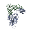
| ||||||||
|---|---|---|---|---|---|---|---|---|---|
| 1 |
| ||||||||
| Unit cell |
|
-Components
| #1: Protein | Mass: 48506.043 Da / Num. of mol.: 1 / Fragment: mitochondrial PheRS, UNP residues 38-451 Source method: isolated from a genetically manipulated source Source: (gene. exp.) Homo sapiens (human) / Gene: FARS2, FARS1, HSPC320 / Production host: Escherichia coli (E. coli) / References: UniProt: O95363, phenylalanine-tRNA ligase |
|---|---|
| #2: RNA chain | Mass: 24486.523 Da / Num. of mol.: 1 Source method: isolated from a genetically manipulated source Source: (gene. exp.) Thermus thermophilus (bacteria) / Production host: Thermus (bacteria) |
-Experimental details
-Experiment
| Experiment | Method: X-RAY DIFFRACTION / Number of used crystals: 1 |
|---|
- Sample preparation
| Crystal | Density Matthews: 3.28 Å3/Da / Density % sol: 62.53 % |
|---|---|
| Crystal grow | Temperature: 292 K / Method: small tubes / pH: 7 Details: 0.2 M ammonium sulfate, 100 mM MES mono-hydrate, pH 7, and 30 % w/v PEG-MME 5,000, SMALL TUBES, temperature 292K |
-Data collection
| Diffraction | Mean temperature: 100 K | |||||||||||||||||||||
|---|---|---|---|---|---|---|---|---|---|---|---|---|---|---|---|---|---|---|---|---|---|---|
| Diffraction source | Source: SYNCHROTRON / Site: ESRF  / Beamline: ID14-4 / Wavelength: 0.93 Å / Beamline: ID14-4 / Wavelength: 0.93 Å | |||||||||||||||||||||
| Detector | Type: ADSC QUANTUM 315r / Detector: CCD / Date: Apr 12, 2010 | |||||||||||||||||||||
| Radiation | Monochromator: GRAPHITE / Protocol: SINGLE WAVELENGTH / Monochromatic (M) / Laue (L): M / Scattering type: x-ray | |||||||||||||||||||||
| Radiation wavelength | Wavelength: 0.93 Å / Relative weight: 1 | |||||||||||||||||||||
| Reflection | Resolution: 3.05→38.95 Å / Num. all: 18827 / Num. obs: 18827 / % possible obs: 99 % / Observed criterion σ(F): 1 / Observed criterion σ(I): 1 | |||||||||||||||||||||
| Reflection shell |
|
- Processing
| Software |
| ||||||||||||||||||||
|---|---|---|---|---|---|---|---|---|---|---|---|---|---|---|---|---|---|---|---|---|---|
| Refinement | Method to determine structure: MOLECULAR REPLACEMENT / Resolution: 3.05→38.94 Å / σ(F): 2 / Stereochemistry target values: Engh & Huber
| ||||||||||||||||||||
| Refinement step | Cycle: LAST / Resolution: 3.05→38.94 Å
| ||||||||||||||||||||
| Refine LS restraints | Type: f_angle_d / Dev ideal: 1.54 |
