Mass: 27478.965 Da / Num. of mol.: 16 Source method: isolated from a genetically manipulated source Source: (gene. exp.) Hyphomicrobium sp. (strain MC1) (bacteria) Gene: HYPMC_4374 / Production host: Escherichia coli BL21(DE3) (bacteria) / References: UniProt: F8JB59
-
Experimental details
-
Experiment
Experiment
Method: X-RAY DIFFRACTION / Number of used crystals: 1
-
Sample preparation
Crystal
Density Matthews: 3.55 Å3/Da / Density % sol: 65.35 %
Crystal grow
Temperature: 298.15 K / Method: vapor diffusion, sitting drop / pH: 8.5 Details: 0.01 M cobalt(II) chloride, 0.1 M Tris, 20 % PVP K15. The protein crystallized in presence of the proteasome inhibitor bortezomib.
Resolution: 3.2→20 Å / Cor.coef. Fo:Fc: 0.938 / Cor.coef. Fo:Fc free: 0.906 / SU B: 28.845 / SU ML: 0.464 / Cross valid method: THROUGHOUT / ESU R Free: 0.514 / Details: HYDROGENS HAVE BEEN ADDED IN THE RIDING POSITIONS
Rfactor
Num. reflection
% reflection
Selection details
Rfree
0.2787
4957
5 %
RANDOM
Rwork
0.23502
-
-
-
obs
0.23715
94184
98.4 %
-
Solvent computation
Ion probe radii: 0.8 Å / Shrinkage radii: 0.8 Å / VDW probe radii: 1.2 Å
 Movie
Movie Controller


 Open data
 Basic information
 Components
 Keywords
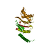
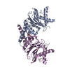
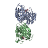
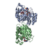
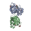
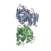
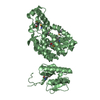
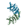
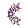
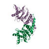
 HYDROLASE / Ntn-hydrolase-fold /
  Function and homology information
 Authors
 Germany, 1items
  Citation
 Structure visualization
 Downloads & links
 Other downloads
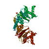


 PDBj
PDBj


 Assembly


 Hyphomicrobium sp. (strain MC1) (bacteria)
 Sample preparation
 / Beamline: X06SA / Wavelength: 1 Å
 Processing