Entry Database : PDB / ID : 5nypTitle Anbu from Hyphomicrobium sp. strain MC1 - SG: R32 Anbu Keywords / / / Function / homology / / / / / / / Biological species Hyphomicrobium sp. Method / / / Resolution : 1.9 Å Authors Vielberg, M.-T. / Groll, M. Funding support Organization Grant number Country German Research Foundation SFB1035
Journal : J. Mol. Biol. / Year : 2018Title : On the Trails of the Proteasome Fold: Structural and Functional Analysis of the Ancestral beta-Subunit Protein Anbu.Authors : Vielberg, M.T. / Bauer, V.C. / Groll, M. History Deposition May 11, 2017 Deposition site / Processing site Revision 1.0 May 24, 2017 Provider / Type Revision 1.1 Feb 28, 2018 Group / Database references / Category / citation_author / pdbx_audit_supportItem _citation.country / _citation.journal_abbrev ... _citation.country / _citation.journal_abbrev / _citation.journal_id_ASTM / _citation.journal_id_CSD / _citation.journal_id_ISSN / _citation.pdbx_database_id_DOI / _citation.pdbx_database_id_PubMed / _citation.title / _citation.year / _citation_author.name / _pdbx_audit_support.funding_organization Revision 1.2 Mar 7, 2018 Group / Category Item / _citation.page_first / _citation.page_lastRevision 1.3 Jan 17, 2024 Group Data collection / Database references ... Data collection / Database references / Derived calculations / Refinement description Category chem_comp_atom / chem_comp_bond ... chem_comp_atom / chem_comp_bond / database_2 / pdbx_initial_refinement_model / pdbx_struct_conn_angle / struct_conn Item _database_2.pdbx_DOI / _database_2.pdbx_database_accession ... _database_2.pdbx_DOI / _database_2.pdbx_database_accession / _pdbx_struct_conn_angle.ptnr1_auth_seq_id / _pdbx_struct_conn_angle.ptnr1_symmetry / _pdbx_struct_conn_angle.ptnr3_auth_seq_id / _pdbx_struct_conn_angle.ptnr3_symmetry / _pdbx_struct_conn_angle.value / _struct_conn.pdbx_dist_value / _struct_conn.ptnr2_auth_seq_id / _struct_conn.ptnr2_symmetry
Show all Show less
 Movie
Movie Controller


 Open data
 Basic information
 Components
 Keywords
 HYDROLASE / Ntn-hydrolase-fold /
  Function and homology information
 Authors
 Germany, 1items
  Citation
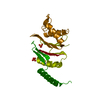
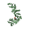
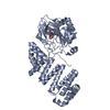
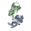
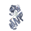
 Structure visualization
 Downloads & links
 Other downloads
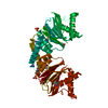


 PDBj
PDBj


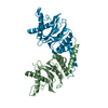
 Assembly
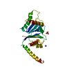

 Hyphomicrobium sp. (strain MC1) (bacteria)
 Mass: 22.990 Da / Num. of mol.: 1 / Source method: obtained synthetically / Formula: Na
 Mass: 94.971 Da / Num. of mol.: 2 / Source method: obtained synthetically / Formula: PO4
 Mass: 18.015 Da / Num. of mol.: 56 / Source method: isolated from a natural source / Formula: H2O
 Sample preparation
 / Beamline: ID23-1 / Wavelength: 1 Å
 Processing