 Movie
Movie Controller

[English] 日本語
 Yorodumi
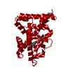
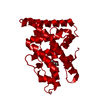
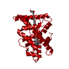
- PDB-1s9p: crystal structure of the ligand-binding domain of the estrogen-re... -
+ Open data

- Basic information
| Entry | Database: PDB / ID: 1s9p | ||||||
|---|---|---|---|---|---|---|---|
| Title | crystal structure of the ligand-binding domain of the estrogen-related receptor gamma in complex with diethylstilbestrol | ||||||
 Components Components | Estrogen-related receptor gamma | ||||||
 Keywords Keywords | TRANSCRIPTION / ligand-binding domain / antagonist complex | ||||||
| Function / homology |  Function and homology information Function and homology informationNuclear Receptor transcription pathway / nuclear steroid receptor activity / retinoic acid receptor signaling pathway / estrogen response element binding / intracellular steroid hormone receptor signaling pathway / steroid binding / RNA polymerase II transcription regulatory region sequence-specific DNA binding / nuclear receptor activity / sequence-specific double-stranded DNA binding / positive regulation of cold-induced thermogenesis ...Nuclear Receptor transcription pathway / nuclear steroid receptor activity / retinoic acid receptor signaling pathway / estrogen response element binding / intracellular steroid hormone receptor signaling pathway / steroid binding / RNA polymerase II transcription regulatory region sequence-specific DNA binding / nuclear receptor activity / sequence-specific double-stranded DNA binding / positive regulation of cold-induced thermogenesis / DNA-binding transcription activator activity, RNA polymerase II-specific / calmodulin binding / regulation of DNA-templated transcription / regulation of transcription by RNA polymerase II / positive regulation of DNA-templated transcription / positive regulation of transcription by RNA polymerase II / DNA binding / zinc ion binding / identical protein binding / nucleusSimilarity search - Function | ||||||
| Biological species |  Mus musculus (house mouse) Mus musculus (house mouse) | ||||||
| Method | X-RAY DIFFRACTION / SYNCHROTRON / MOLECULAR REPLACEMENT / Resolution: 2.13 Å | ||||||
 Authors Authors | Greschik, H. / Flaig, R. / Renaud, J.P. / Moras, D. | ||||||
 Citation Citation | Journal: J.Biol.Chem. / Year: 2004 Title: Structural Basis for the Deactivation of the Estrogen-related Receptor {gamma} by Diethylstilbestrol or 4-Hydroxytamoxifen and Determinants of Selectivity. Authors: Greschik, H. / Flaig, R. / Renaud, J.P. / Moras, D. | ||||||
| History |
|
- Structure visualization
| Structure viewer | Molecule: MolmilJmol/JSmol |
|---|
- Downloads & links
-Download
| PDBx/mmCIF format | 1s9p.cif.gz | 180.1 KB | Display | PDBx/mmCIF format |
|---|---|---|---|---|
| PDB format | pdb1s9p.ent.gz | 146.1 KB | Display | PDB format |
| PDBx/mmJSON format | 1s9p.json.gz | Tree view | PDBx/mmJSON format | |
| Others |  Other downloads Other downloads |
-Validation report
| Arichive directory | https://data.pdbj.org/pub/pdb/validation_reports/s9/1s9pftp://data.pdbj.org/pub/pdb/validation_reports/s9/1s9p | HTTPS FTP |
|---|
-Related structure data
| Related structure data |  1s9qC 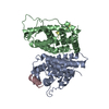 1tfcC  1vjbC 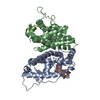 1kv6S S: Starting model for refinement C: citing same article ( |
|---|---|
| Similar structure data |


-Links
 PDBj
PDBj

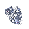
- Assembly
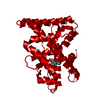
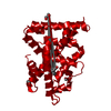
| Deposited unit | 
| ||||||||
|---|---|---|---|---|---|---|---|---|---|
| 1 | 
| ||||||||
| 2 | 
| ||||||||
| 3 | 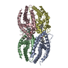
| ||||||||
| 4 | 
| ||||||||
| 5 | 
| ||||||||
| Unit cell |
|
-Components
| #1: Protein | / Estrogen receptor related protein 3 / ERR gamma-2 Mass: 25787.002 Da / Num. of mol.: 4 Source method: isolated from a genetically manipulated source Source: (gene. exp.) Mus musculus (house mouse) / Gene: ESRRG, NR3B3, ERRG2, ERR3, KIAA0832 / Plasmid: pET-15b / Species (production host): Escherichia coli / Production host:  Escherichia coli BL21(DE3) (bacteria) / Strain (production host): BL21(DE3) / References: UniProt: P62509 Escherichia coli BL21(DE3) (bacteria) / Strain (production host): BL21(DE3) / References: UniProt: P62509#2: Chemical | ChemComp-DES / Diethylstilbestrol  Mass: 268.350 Da / Num. of mol.: 4 / Source method: obtained synthetically / Formula: C18H20O2 / Comment: medication, hormone*YM Mass: 268.350 Da / Num. of mol.: 4 / Source method: obtained synthetically / Formula: C18H20O2 / Comment: medication, hormone*YM#3: Water | ChemComp-HOH / | Water Mass: 18.015 Da / Num. of mol.: 206 / Source method: isolated from a natural source / Formula: H2O Mass: 18.015 Da / Num. of mol.: 206 / Source method: isolated from a natural source / Formula: H2O |
|---|
-Experimental details
-Experiment
| Experiment | Method: X-RAY DIFFRACTION / Number of used crystals: 1 |
|---|
- Sample preparation
| Crystal | Density Matthews: 2.57 Å3/Da / Density % sol: 52.06 % |
|---|---|
| Crystal grow | Temperature: 297 K / pH: 8 Details: PEG3350, NaOAc, pH 8.0, VAPOR DIFFUSION, SITTING DROP, temperature 297K, pH 8.00 |
-Data collection
| Diffraction | Mean temperature: 100 K |
|---|---|
| Diffraction source | Source: SYNCHROTRON / Site: ESRF  / Beamline: ID14-1 / Wavelength: 0.934 / Beamline: ID14-1 / Wavelength: 0.934 |
| Detector | Type: ADSC QUANTUM 4 / Detector: CCD / Date: Oct 7, 2002 / Details: SAGITALLY FOCUSING GE(220) AND A MULTILAYER |
| Radiation | Monochromator: DIAMOND (111), GE(220) / Protocol: SINGLE WAVELENGTH / Monochromatic (M) / Laue (L): M / Scattering type: x-ray |
| Radiation wavelength | Wavelength: 0.934 Å / Relative weight: 1 |
| Reflection | Resolution: 2.13→45 Å / Num. obs: 57942 / % possible obs: 99 % / Biso Wilson estimate: 35.1 Å2 / Rsym value: 0.039 / Net I/σ(I): 46.6 |
| Reflection shell | Resolution: 2.13→2.21 Å / Mean I/σ(I) obs: 9.7 / Rsym value: 0.159 / % possible all: 99.8 |
- Processing
| Software |
| ||||||||||||||||||||||||||||||||||||||||||||||||||||||||||||
|---|---|---|---|---|---|---|---|---|---|---|---|---|---|---|---|---|---|---|---|---|---|---|---|---|---|---|---|---|---|---|---|---|---|---|---|---|---|---|---|---|---|---|---|---|---|---|---|---|---|---|---|---|---|---|---|---|---|---|---|---|---|
| Refinement | Method to determine structure: MOLECULAR REPLACEMENT Starting model: PDB ENTRY 1KV6 Resolution: 2.13→29.3 Å / Rfactor Rfree error: 0.005 / Data cutoff high absF: 768655.75 / Data cutoff high rms absF: 768655.75 / Data cutoff low absF: 0 / Isotropic thermal model: RESTRAINED / Cross valid method: THROUGHOUT / σ(F): 2 / Stereochemistry target values: ENGH & HUBER
| ||||||||||||||||||||||||||||||||||||||||||||||||||||||||||||
| Solvent computation | Solvent model: FLAT MODEL / Bsol: 50.1 Å2 / ksol: 0.36 e/Å3 | ||||||||||||||||||||||||||||||||||||||||||||||||||||||||||||
| Displacement parameters | Biso mean: 43.9 Å2
| ||||||||||||||||||||||||||||||||||||||||||||||||||||||||||||
| Refine analyze |
| ||||||||||||||||||||||||||||||||||||||||||||||||||||||||||||
| Refinement step | Cycle: LAST / Resolution: 2.13→29.3 Å
| ||||||||||||||||||||||||||||||||||||||||||||||||||||||||||||
| Refine LS restraints |
| ||||||||||||||||||||||||||||||||||||||||||||||||||||||||||||
| LS refinement shell | Resolution: 2.13→2.21 Å / Rfactor Rfree error: 0.017 / Total num. of bins used: 10
| ||||||||||||||||||||||||||||||||||||||||||||||||||||||||||||
| Xplor file |
|
