 Movie
Movie Controller

[English] 日本語
 Yorodumi
- PDB-3b3x: Crystal structure of class A beta-lactamase of Bacillus lichenifo... -
+ Open data

- Basic information
| Entry | Database: PDB / ID: 3b3x | ||||||
|---|---|---|---|---|---|---|---|
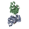
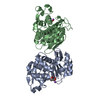
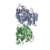
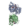
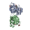
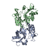
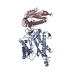
| Title | Crystal structure of class A beta-lactamase of Bacillus licheniformis BS3 with aminocitrate | ||||||
 Components Components | Beta-lactamase | ||||||
 Keywords Keywords | HYDROLASE/HYDROLASE INHIBITOR / beta-lactamase / aminocitrate / Antibiotic resistance / HYDROLASE-HYDROLASE INHIBITOR complex | ||||||
| Function / homology |  Function and homology information Function and homology informationbeta-lactam antibiotic catabolic process / beta-lactamase activity / beta-lactamase / response to antibiotic / plasma membraneSimilarity search - Function | ||||||
| Biological species |  Bacillus licheniformis (bacteria) Bacillus licheniformis (bacteria) | ||||||
| Method | X-RAY DIFFRACTION / MOLECULAR REPLACEMENT / Resolution: 2.5 Å | ||||||
 Authors Authors | Sauvage, E. / Herman, R. / Kerff, F. / Charlier, P. | ||||||
 Citation Citation | Journal: Bioorg.Med.Chem.Lett. / Year: 2008 Title: 2-Aminopropane-1,2,3-tricarboxylic acid: Synthesis and co-crystallization with the class A beta-lactamase BS3 of Bacillus licheniformis Authors: Beck, J. / Sauvage, E. / Charlier, P. / Marchand-Brynaert, J. | ||||||
| History |
|
- Structure visualization
| Structure viewer | Molecule: MolmilJmol/JSmol |
|---|
- Downloads & links
-Download
| PDBx/mmCIF format | 3b3x.cif.gz | 113.7 KB | Display | PDBx/mmCIF format |
|---|---|---|---|---|
| PDB format | pdb3b3x.ent.gz | 88.7 KB | Display | PDB format |
| PDBx/mmJSON format | 3b3x.json.gz | Tree view | PDBx/mmJSON format | |
| Others |  Other downloads Other downloads |
-Validation report
| Arichive directory | https://data.pdbj.org/pub/pdb/validation_reports/b3/3b3xftp://data.pdbj.org/pub/pdb/validation_reports/b3/3b3x | HTTPS FTP |
|---|
-Related structure data
| Related structure data |  1w7fS S: Starting model for refinement |
|---|---|
| Similar structure data |


-Links
 PDBj
PDBj


- Assembly
| Deposited unit | 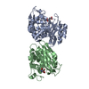
| ||||||||
|---|---|---|---|---|---|---|---|---|---|
| 1 |
| ||||||||
| Unit cell |
|
-Components
| #1: Protein | Mass: 29513.303 Da / Num. of mol.: 2 / Source method: isolated from a natural source / Source: (natural) Bacillus licheniformis (bacteria) / Strain: BS3References: UniProt: P94458, UniProt: P00808*PLUS, beta-lactamase#2: Chemical |   Mass: 191.139 Da / Num. of mol.: 2 / Source method: obtained synthetically / Formula: C6H9NO6 Mass: 191.139 Da / Num. of mol.: 2 / Source method: obtained synthetically / Formula: C6H9NO6#3: Water | ChemComp-HOH / | Water Mass: 18.015 Da / Num. of mol.: 156 / Source method: isolated from a natural source / Formula: H2O Mass: 18.015 Da / Num. of mol.: 156 / Source method: isolated from a natural source / Formula: H2O |
|---|
-Experimental details
-Experiment
| Experiment | Method: X-RAY DIFFRACTION / Number of used crystals: 1 |
|---|
- Sample preparation
| Crystal | Density Matthews: 2.64 Å3/Da / Density % sol: 53.37 % |
|---|---|
| Crystal grow | Temperature: 293 K / Method: hanging drop / pH: 7.2 Details: 5micro-l of a protein solution (at a concentration of 38mg/ml in 50mM NaCl, 10mM Tris buffer, pH 7.2), 4micro-l of 8% PEG 6000 in 100mM sodium aminocitrate buffer (pH 3.4) plus 1micro-l of 0. ...Details: 5micro-l of a protein solution (at a concentration of 38mg/ml in 50mM NaCl, 10mM Tris buffer, pH 7.2), 4micro-l of 8% PEG 6000 in 100mM sodium aminocitrate buffer (pH 3.4) plus 1micro-l of 0.1M urea additive, equilibrated against 1ml of a 20% PEG 6000, hanging drop, temperature 293K |
-Data collection
| Diffraction | Mean temperature: 100 K | ||||||||||||||||||||||||||||||||||||||||||||||||||||||||||||||||||||||||||||||||||||||||
|---|---|---|---|---|---|---|---|---|---|---|---|---|---|---|---|---|---|---|---|---|---|---|---|---|---|---|---|---|---|---|---|---|---|---|---|---|---|---|---|---|---|---|---|---|---|---|---|---|---|---|---|---|---|---|---|---|---|---|---|---|---|---|---|---|---|---|---|---|---|---|---|---|---|---|---|---|---|---|---|---|---|---|---|---|---|---|---|---|---|
| Diffraction source | Source: ROTATING ANODE / Type: RIGAKU RU200 / Wavelength: 1.5418 Å | ||||||||||||||||||||||||||||||||||||||||||||||||||||||||||||||||||||||||||||||||||||||||
| Detector | Type: MAR scanner 345 mm plate / Detector: IMAGE PLATE / Date: Mar 4, 2005 / Details: mirrors | ||||||||||||||||||||||||||||||||||||||||||||||||||||||||||||||||||||||||||||||||||||||||
| Radiation | Protocol: SINGLE WAVELENGTH / Monochromatic (M) / Laue (L): M / Scattering type: x-ray | ||||||||||||||||||||||||||||||||||||||||||||||||||||||||||||||||||||||||||||||||||||||||
| Radiation wavelength | Wavelength: 1.5418 Å / Relative weight: 1 | ||||||||||||||||||||||||||||||||||||||||||||||||||||||||||||||||||||||||||||||||||||||||
| Reflection | Resolution: 2.5→63.758 Å / Num. obs: 17440 / % possible obs: 82.3 % / Redundancy: 1.9 % / Rmerge(I) obs: 0.083 / Rsym value: 0.083 / Net I/σ(I): 7.6 | ||||||||||||||||||||||||||||||||||||||||||||||||||||||||||||||||||||||||||||||||||||||||
| Reflection shell |
|
- Processing
| Software |
| ||||||||||||||||||||||||||||||||||||||||||||||||||||||||||||||||||||||||||||||||||||||||||
|---|---|---|---|---|---|---|---|---|---|---|---|---|---|---|---|---|---|---|---|---|---|---|---|---|---|---|---|---|---|---|---|---|---|---|---|---|---|---|---|---|---|---|---|---|---|---|---|---|---|---|---|---|---|---|---|---|---|---|---|---|---|---|---|---|---|---|---|---|---|---|---|---|---|---|---|---|---|---|---|---|---|---|---|---|---|---|---|---|---|---|---|
| Refinement | Method to determine structure: MOLECULAR REPLACEMENT Starting model: PDB ENTRY 1W7F Resolution: 2.5→36.47 Å / Cor.coef. Fo:Fc: 0.9 / Cor.coef. Fo:Fc free: 0.831 / SU B: 11.704 / SU ML: 0.261 / Cross valid method: THROUGHOUT / σ(F): 0 / ESU R Free: 0.393 / Stereochemistry target values: MAXIMUM LIKELIHOOD / Details: HYDROGENS HAVE BEEN ADDED IN THE RIDING POSITIONS
| ||||||||||||||||||||||||||||||||||||||||||||||||||||||||||||||||||||||||||||||||||||||||||
| Solvent computation | Ion probe radii: 0.8 Å / Shrinkage radii: 0.8 Å / VDW probe radii: 1.4 Å / Solvent model: MASK | ||||||||||||||||||||||||||||||||||||||||||||||||||||||||||||||||||||||||||||||||||||||||||
| Displacement parameters | Biso mean: 22.959 Å2
| ||||||||||||||||||||||||||||||||||||||||||||||||||||||||||||||||||||||||||||||||||||||||||
| Refinement step | Cycle: LAST / Resolution: 2.5→36.47 Å
| ||||||||||||||||||||||||||||||||||||||||||||||||||||||||||||||||||||||||||||||||||||||||||
| Refine LS restraints |
| ||||||||||||||||||||||||||||||||||||||||||||||||||||||||||||||||||||||||||||||||||||||||||
| LS refinement shell | Resolution: 2.5→2.565 Å / Total num. of bins used: 20
|
