 Movie
Movie Controller


+ Open data

- Basic information
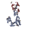
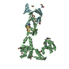
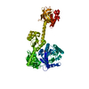
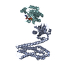
| Entry | Database: PDB / ID: 5msm | ||||||||||||
|---|---|---|---|---|---|---|---|---|---|---|---|---|---|
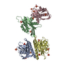
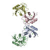
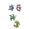
| Title | Structure of the Dcc1-Ctf8-Ctf18C Trimer | ||||||||||||
 Components Components |
| ||||||||||||
 Keywords Keywords |  CELL CYCLE / winged-helix / DNA repair CELL CYCLE / winged-helix / DNA repair | ||||||||||||
| Function / homology |  Function and homology information Function and homology informationmaintenance of mitotic sister chromatid cohesion / Ctf18 RFC-like complex / maintenance of DNA trinucleotide repeats / mitotic sister chromatid cohesion / nuclear replication fork / chromosome, centromeric region / DNA replication initiation / double-strand break repair via homologous recombination / DNA replication / chromatin ...maintenance of mitotic sister chromatid cohesion / Ctf18 RFC-like complex / maintenance of DNA trinucleotide repeats / mitotic sister chromatid cohesion / nuclear replication fork / chromosome, centromeric region / DNA replication initiation / double-strand break repair via homologous recombination / DNA replication / chromatin / ATP hydrolysis activity / mitochondrion / DNA binding / ATP binding / nucleusSimilarity search - Function | ||||||||||||
| Biological species |  Saccharomyces cerevisiae S288c (yeast) Saccharomyces cerevisiae S288c (yeast) | ||||||||||||
| Method | X-RAY DIFFRACTION / SYNCHROTRON / MOLECULAR REPLACEMENT / Resolution: 2.29 Å | ||||||||||||
 Authors Authors | Wade, B.O. / Singleton, M.R. | ||||||||||||
| Funding support |  United Kingdom, 3items United Kingdom, 3items
| ||||||||||||
 Citation Citation | Journal: EMBO Rep. / Year: 2017 Title: Structural studies of RFC(C)(tf18) reveal a novel chromatin recruitment role for Dcc1. Authors: Wade, B.O. / Liu, H.W. / Samora, C.P. / Uhlmann, F. / Singleton, M.R. | ||||||||||||
| History |
|
- Structure visualization
| Structure viewer | Molecule: MolmilJmol/JSmol |
|---|
- Downloads & links
-Download
| PDBx/mmCIF format | 5msm.cif.gz | 226.8 KB | Display | PDBx/mmCIF format |
|---|---|---|---|---|
| PDB format | pdb5msm.ent.gz | 181 KB | Display | PDB format |
| PDBx/mmJSON format | 5msm.json.gz | Tree view | PDBx/mmJSON format | |
| Others |  Other downloads Other downloads |
-Validation report
| Arichive directory | https://data.pdbj.org/pub/pdb/validation_reports/ms/5msmftp://data.pdbj.org/pub/pdb/validation_reports/ms/5msm | HTTPS FTP |
|---|
-Related structure data
| Related structure data |  5msnSC S: Starting model for refinement C: citing same article ( |
|---|---|
| Similar structure data |

-Links
 PDBj
PDBj- Assembly
| Deposited unit | 
| ||||||||
|---|---|---|---|---|---|---|---|---|---|
| 1 | 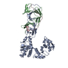
| ||||||||
| 2 | 
| ||||||||
| Unit cell |
|
-Components
| #1: Protein | Mass: 44133.785 Da / Num. of mol.: 2 Source method: isolated from a genetically manipulated source Source: (gene. exp.) Saccharomyces cerevisiae S288c (yeast) / Gene: DCC1, YCL016C, YCL16C / Production host:  Escherichia coli (E. coli) / References: UniProt: P25559 Escherichia coli (E. coli) / References: UniProt: P25559#2: Protein | Mass: 15189.688 Da / Num. of mol.: 2 Source method: isolated from a genetically manipulated source Source: (gene. exp.) Saccharomyces cerevisiae S288c (yeast) / Gene: CTF8, YHR191C / Production host: Escherichia coli (E. coli) / References: UniProt: P38877#3: Protein | Mass: 8915.728 Da / Num. of mol.: 2 Source method: isolated from a genetically manipulated source Source: (gene. exp.) Saccharomyces cerevisiae S288c (yeast) / Gene: CTF18, CHL12, YMR078C, YM9582.03C / Production host: Escherichia coli (E. coli) / References: UniProt: P49956#4: Water | ChemComp-HOH / | Water Mass: 18.015 Da / Num. of mol.: 266 / Source method: isolated from a natural source / Formula: H2O Mass: 18.015 Da / Num. of mol.: 266 / Source method: isolated from a natural source / Formula: H2O |
|---|
-Experimental details
-Experiment
| Experiment | Method: X-RAY DIFFRACTION / Number of used crystals: 1 |
|---|
- Sample preparation
| Crystal | Density Matthews: 2.14 Å3/Da / Density % sol: 42.47 % |
|---|---|
| Crystal grow | Temperature: 293 K / Method: vapor diffusion, sitting drop Details: 0.1M Bis-Tris Propane pH 6.3, 0.2M NaBr and 17% PEG 3,350 |
-Data collection
| Diffraction | Mean temperature: 93 K |
|---|---|
| Diffraction source | Source: SYNCHROTRON / Site: Diamond / Beamline: I03 / Wavelength: 0.9763 Å |
| Detector | Type: DECTRIS PILATUS3 6M / Detector: PIXEL / Date: Jul 26, 2015 |
| Radiation | Protocol: SINGLE WAVELENGTH / Monochromatic (M) / Laue (L): M / Scattering type: x-ray |
| Radiation wavelength | Wavelength: 0.9763 Å / Relative weight: 1 |
| Reflection | Resolution: 2.29→60.64 Å / Num. obs: 46232 / % possible obs: 90 % / Redundancy: 3 % / Rmerge(I) obs: 0.08522 / Net I/σ(I): 7.15 |
| Reflection shell | Resolution: 2.29→2.372 Å / Redundancy: 1.9 % / Rmerge(I) obs: 0.4557 / Mean I/σ(I) obs: 1.31 / Num. unique all: 2926 / % possible all: 57 |
- Processing
| Software |
| |||||||||||||||||||||||||||||||||||||||||||||||||||||||||||||||||||||||||||||||||||||||||||||||||||||||||||||||||||||||
|---|---|---|---|---|---|---|---|---|---|---|---|---|---|---|---|---|---|---|---|---|---|---|---|---|---|---|---|---|---|---|---|---|---|---|---|---|---|---|---|---|---|---|---|---|---|---|---|---|---|---|---|---|---|---|---|---|---|---|---|---|---|---|---|---|---|---|---|---|---|---|---|---|---|---|---|---|---|---|---|---|---|---|---|---|---|---|---|---|---|---|---|---|---|---|---|---|---|---|---|---|---|---|---|---|---|---|---|---|---|---|---|---|---|---|---|---|---|---|---|---|
| Refinement | Method to determine structure: MOLECULAR REPLACEMENT Starting model: 5MSN Resolution: 2.29→60.64 Å / SU ML: 0.33 / Cross valid method: FREE R-VALUE / σ(F): 1.34 / Phase error: 32.12
| |||||||||||||||||||||||||||||||||||||||||||||||||||||||||||||||||||||||||||||||||||||||||||||||||||||||||||||||||||||||
| Solvent computation | Shrinkage radii: 0.9 Å / VDW probe radii: 1.11 Å | |||||||||||||||||||||||||||||||||||||||||||||||||||||||||||||||||||||||||||||||||||||||||||||||||||||||||||||||||||||||
| Refinement step | Cycle: LAST / Resolution: 2.29→60.64 Å
| |||||||||||||||||||||||||||||||||||||||||||||||||||||||||||||||||||||||||||||||||||||||||||||||||||||||||||||||||||||||
| Refine LS restraints |
| |||||||||||||||||||||||||||||||||||||||||||||||||||||||||||||||||||||||||||||||||||||||||||||||||||||||||||||||||||||||
| LS refinement shell |
|
