 Movie
Movie Controller

[English] 日本語
 Yorodumi
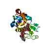
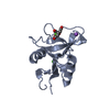
- PDB-2ox8: Human Scavenger Receptor C-type Lectin carbohydrate-recognition d... -
+ Open data

- Basic information
| Entry | Database: PDB / ID: 2ox8 | ||||||
|---|---|---|---|---|---|---|---|
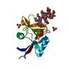
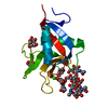
| Title | Human Scavenger Receptor C-type Lectin carbohydrate-recognition domain. | ||||||
 Components Components | Scavenger receptor with C-type lectin type I | ||||||
 Keywords Keywords | SUGAR BINDING PROTEIN /  c-type lectin c-type lectin | ||||||
| Function / homology |  Function and homology information Function and homology informationcarbohydrate mediated signaling / positive regulation of cell adhesion molecule production / galactose binding / toll-like receptor 3 signaling pathway / low-density lipoprotein particle binding / scavenger receptor activity / phagocytosis, recognition / collagen trimer / Scavenging by Class A Receptors / pattern recognition receptor activity ...carbohydrate mediated signaling / positive regulation of cell adhesion molecule production / galactose binding / toll-like receptor 3 signaling pathway / low-density lipoprotein particle binding / scavenger receptor activity / phagocytosis, recognition / collagen trimer / Scavenging by Class A Receptors / pattern recognition receptor activity / cellular response to exogenous dsRNA / regulation of immune response / receptor-mediated endocytosis / extracellular matrix / defense response / endocytic vesicle membrane / Immunoregulatory interactions between a Lymphoid and a non-Lymphoid cell / membrane => GO:0016020 / defense response to bacterium / innate immune response / extracellular space / metal ion binding / plasma membraneSimilarity search - Function | ||||||
| Biological species |  Homo sapiens (human) Homo sapiens (human) | ||||||
| Method | X-RAY DIFFRACTION / SYNCHROTRON / MOLECULAR REPLACEMENT / Resolution: 2.5 Å | ||||||
 Authors Authors | Weis, W.I. / Feinberg, H. / Drickamer, K. / Taylor, M.E. | ||||||
 Citation Citation | Journal: J.Biol.Chem. / Year: 2007 Title: Scavenger receptor C-type lectin binds to the leukocyte cell surface glycan Lewis(x) by a novel mechanism. Authors: Feinberg, H. / Taylor, M.E. / Weis, W.I. | ||||||
| History |
|
- Structure visualization
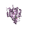
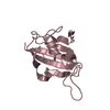
| Structure viewer | Molecule: MolmilJmol/JSmol |
|---|
- Downloads & links
-Download
| PDBx/mmCIF format | 2ox8.cif.gz | 119 KB | Display | PDBx/mmCIF format |
|---|---|---|---|---|
| PDB format | pdb2ox8.ent.gz | 98.2 KB | Display | PDB format |
| PDBx/mmJSON format | 2ox8.json.gz | Tree view | PDBx/mmJSON format | |
| Others |  Other downloads Other downloads |
-Validation report
| Arichive directory | https://data.pdbj.org/pub/pdb/validation_reports/ox/2ox8ftp://data.pdbj.org/pub/pdb/validation_reports/ox/2ox8 | HTTPS FTP |
|---|
-Related structure data


-Links
 PDBj
PDBj


- Assembly
| Deposited unit | 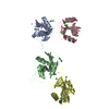
| ||||||||
|---|---|---|---|---|---|---|---|---|---|
| 1 | 
| ||||||||
| 2 | 
| ||||||||
| 3 | 
| ||||||||
| 4 | 
| ||||||||
| Unit cell |
|
-Components
| #1: Protein | Mass: 16462.012 Da / Num. of mol.: 4 / Fragment: CRD domain Source method: isolated from a genetically manipulated source Source: (gene. exp.) Homo sapiens (human) / Gene: SRCL / Production host:  Escherichia coli (E. coli) / References: UniProt: Q9BYH7, UniProt: Q5KU26*PLUS Escherichia coli (E. coli) / References: UniProt: Q9BYH7, UniProt: Q5KU26*PLUS#2: Chemical | ChemComp-ZN /   Mass: 65.409 Da / Num. of mol.: 16 / Source method: obtained synthetically / Formula: Zn Mass: 65.409 Da / Num. of mol.: 16 / Source method: obtained synthetically / Formula: Zn#3: Chemical | ChemComp-CL / Chloride  Mass: 35.453 Da / Num. of mol.: 12 / Source method: obtained synthetically / Formula: Cl Mass: 35.453 Da / Num. of mol.: 12 / Source method: obtained synthetically / Formula: Cl#4: Chemical | ChemComp-CA /   Mass: 40.078 Da / Num. of mol.: 6 / Source method: obtained synthetically / Formula: Ca Mass: 40.078 Da / Num. of mol.: 6 / Source method: obtained synthetically / Formula: Ca#5: Water | ChemComp-HOH / | Water Mass: 18.015 Da / Num. of mol.: 33 / Source method: isolated from a natural source / Formula: H2O Mass: 18.015 Da / Num. of mol.: 33 / Source method: isolated from a natural source / Formula: H2O |
|---|
-Experimental details
-Experiment
| Experiment | Method: X-RAY DIFFRACTION / Number of used crystals: 1 |
|---|
- Sample preparation
| Crystal | Density Matthews: 1.9 Å3/Da / Density % sol: 35.39 % |
|---|---|
| Crystal grow | Temperature: 298 K / Method: vapor diffusion, hanging drop / pH: 7 Details: The protein solution contained 10mg ml-1 protein, 8mM CaCl2 , 8mM Tris pH=7.8, 20mM NaCl and 10mM Lewisx. The reservoir solution contained 8% Peg 8K, 0.2M ZnAc2 and 0.1 M Tris, pH 7.0. , ...Details: The protein solution contained 10mg ml-1 protein, 8mM CaCl2 , 8mM Tris pH=7.8, 20mM NaCl and 10mM Lewisx. The reservoir solution contained 8% Peg 8K, 0.2M ZnAc2 and 0.1 M Tris, pH 7.0. , VAPOR DIFFUSION, HANGING DROP, temperature 298K |
-Data collection
| Diffraction | Mean temperature: 100 K |
|---|---|
| Diffraction source | Source: SYNCHROTRON / Site: ALS  / Beamline: 8.2.1 / Wavelength: 1 Å / Beamline: 8.2.1 / Wavelength: 1 Å |
| Detector | Type: ADSC QUANTUM 315 / Detector: CCD / Date: Nov 22, 2005 |
| Radiation | Protocol: SINGLE WAVELENGTH / Monochromatic (M) / Laue (L): M / Scattering type: x-ray |
| Radiation wavelength | Wavelength: 1 Å / Relative weight: 1 |
| Reflection | Resolution: 2.5→30.9 Å / Num. obs: 16533 / % possible obs: 98.4 % / Observed criterion σ(F): 0 / Redundancy: 1.9 % / Biso Wilson estimate: 39.9 Å2 / Rsym value: 0.069 |
| Reflection shell | Resolution: 2.5→2.57 Å / Rsym value: 0.24 / % possible all: 99.5 |
- Processing
| Software |
| ||||||||||||||||||||||||||||||||||||||||||||||||||||||||||||||||||||||||||||||||
|---|---|---|---|---|---|---|---|---|---|---|---|---|---|---|---|---|---|---|---|---|---|---|---|---|---|---|---|---|---|---|---|---|---|---|---|---|---|---|---|---|---|---|---|---|---|---|---|---|---|---|---|---|---|---|---|---|---|---|---|---|---|---|---|---|---|---|---|---|---|---|---|---|---|---|---|---|---|---|---|---|---|
| Refinement | Method to determine structure: MOLECULAR REPLACEMENT / Resolution: 2.5→30.92 Å / Rfactor Rfree error: 0.011 / Data cutoff high absF: 709657.69 / Data cutoff low absF: 0 / Isotropic thermal model: RESTRAINED / Cross valid method: THROUGHOUT / σ(F): 0
| ||||||||||||||||||||||||||||||||||||||||||||||||||||||||||||||||||||||||||||||||
| Solvent computation | Solvent model: FLAT MODEL / Bsol: 59.7607 Å2 / ksol: 0.356584 e/Å3 | ||||||||||||||||||||||||||||||||||||||||||||||||||||||||||||||||||||||||||||||||
| Displacement parameters | Biso mean: 31.7 Å2
| ||||||||||||||||||||||||||||||||||||||||||||||||||||||||||||||||||||||||||||||||
| Refine analyze |
| ||||||||||||||||||||||||||||||||||||||||||||||||||||||||||||||||||||||||||||||||
| Refinement step | Cycle: LAST / Resolution: 2.5→30.92 Å
| ||||||||||||||||||||||||||||||||||||||||||||||||||||||||||||||||||||||||||||||||
| Refine LS restraints |
| ||||||||||||||||||||||||||||||||||||||||||||||||||||||||||||||||||||||||||||||||
| LS refinement shell | Resolution: 2.5→2.66 Å / Rfactor Rfree error: 0.032 / Total num. of bins used: 6
| ||||||||||||||||||||||||||||||||||||||||||||||||||||||||||||||||||||||||||||||||
| Xplor file |
|
