 Movie
Movie Controller

[English] 日本語
 Yorodumi
- PDB-5eh1: Crystal structure of the extracellular part of receptor 2 of huma... -
+ Open data

- Basic information
| Entry | Database: PDB / ID: 5eh1 | ||||||
|---|---|---|---|---|---|---|---|
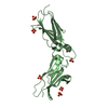
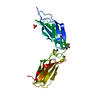
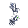
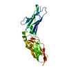
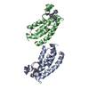
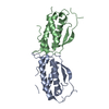
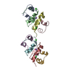
| Title | Crystal structure of the extracellular part of receptor 2 of human interferon gamma | ||||||
 Components Components | Interferon gamma receptor 2 | ||||||
 Keywords Keywords | CYTOKINE / interferon gamma / immunity / fibronectin III domain | ||||||
| Function / homology |  Function and homology information Function and homology informationtype II interferon receptor activity / type III interferon-mediated signaling pathway / cytokine receptor activity / IFNG signaling activates MAPKs / type II interferon-mediated signaling pathway / Regulation of IFNG signaling / microglial cell activation / response to virus / cytoplasmic vesicle membrane / cellular response to virus ...type II interferon receptor activity / type III interferon-mediated signaling pathway / cytokine receptor activity / IFNG signaling activates MAPKs / type II interferon-mediated signaling pathway / Regulation of IFNG signaling / microglial cell activation / response to virus / cytoplasmic vesicle membrane / cellular response to virus / cytokine-mediated signaling pathway / Interferon gamma signaling / defense response to virus / Potential therapeutics for SARS / cell surface receptor signaling pathway / Golgi membrane / endoplasmic reticulum membrane / endoplasmic reticulum / plasma membraneSimilarity search - Function | ||||||
| Biological species |  Homo sapiens (human) Homo sapiens (human) | ||||||
| Method | X-RAY DIFFRACTION / SYNCHROTRON / MOLECULAR REPLACEMENT / Resolution: 1.8 Å | ||||||
 Authors Authors | Kolenko, P. / Mikulecky, P. / Zahradnik, J. / Dohnalek, J. / Koval, T. / Cerny, J. / Necasova, I. / Schneider, B. | ||||||
 Citation Citation | Journal: Acta Crystallogr D Struct Biol / Year: 2016 Title: Crystal structure of human interferon-gamma receptor 2 reveals the structural basis for receptor specificity. Authors: Mikulecky, P. / Zahradnik, J. / Kolenko, P. / Cerny, J. / Charnavets, T. / Kolarova, L. / Necasova, I. / Pham, P.N. / Schneider, B. | ||||||
| History |
|
- Structure visualization
| Structure viewer | Molecule: MolmilJmol/JSmol |
|---|
- Downloads & links
-Download
| PDBx/mmCIF format | 5eh1.cif.gz | 63.2 KB | Display | PDBx/mmCIF format |
|---|---|---|---|---|
| PDB format | pdb5eh1.ent.gz | 48.9 KB | Display | PDB format |
| PDBx/mmJSON format | 5eh1.json.gz | Tree view | PDBx/mmJSON format | |
| Others |  Other downloads Other downloads |
-Validation report
| Arichive directory | https://data.pdbj.org/pub/pdb/validation_reports/eh/5eh1ftp://data.pdbj.org/pub/pdb/validation_reports/eh/5eh1 | HTTPS FTP |
|---|
-Related structure data
| Similar structure data |
|---|


-Links
 PDBj
PDBj


- Assembly
| Deposited unit | 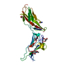
| ||||||||
|---|---|---|---|---|---|---|---|---|---|
| 1 |
| ||||||||
| Unit cell |
| ||||||||
| Components on special symmetry positions |
|
-Components
| #1: Protein | / IFN-gamma-R2 / Interferon gamma receptor accessory factor 1 / AF-1 / Interferon gamma transducer 1 Mass: 26208.461 Da / Num. of mol.: 1 Source method: isolated from a genetically manipulated source Source: (gene. exp.) Homo sapiens (human) / Gene: IFNGR2, IFNGT1 / Plasmid: pMT-BiP-V5-His_ADetails (production host): pMT-BiP-V5-His_A vector co-transfected with the pCoBlast for selection Cell line (production host): Schneider 2 cells / Production host:  Drosophila melanogaster (fruit fly) / References: UniProt: P38484 Drosophila melanogaster (fruit fly) / References: UniProt: P38484 | ||||
|---|---|---|---|---|---|
| #2: Chemical | ChemComp-CYS / Cysteine  Type: L-peptide linking / Mass: 121.158 Da / Num. of mol.: 1 / Source method: obtained synthetically / Formula: C3H7NO2S Type: L-peptide linking / Mass: 121.158 Da / Num. of mol.: 1 / Source method: obtained synthetically / Formula: C3H7NO2S | ||||
| #3: Sugar | N-Acetylglucosamine  Type: D-saccharide, beta linking / Mass: 221.208 Da / Num. of mol.: 3 Type: D-saccharide, beta linking / Mass: 221.208 Da / Num. of mol.: 3Source method: isolated from a genetically manipulated source Formula: C8H15NO6 #4: Chemical | Glycerol  Mass: 92.094 Da / Num. of mol.: 3 / Source method: obtained synthetically / Formula: C3H8O3 Mass: 92.094 Da / Num. of mol.: 3 / Source method: obtained synthetically / Formula: C3H8O3#5: Water | ChemComp-HOH / | Water Mass: 18.015 Da / Num. of mol.: 311 / Source method: isolated from a natural source / Formula: H2O Mass: 18.015 Da / Num. of mol.: 311 / Source method: isolated from a natural source / Formula: H2O |
-Experimental details
-Experiment
| Experiment | Method: X-RAY DIFFRACTION |
|---|
- Sample preparation
| Crystal | Density Matthews: 3.51 Å3/Da / Density % sol: 64.93 % |
|---|---|
| Crystal grow | Temperature: 293 K / Method: vapor diffusion, hanging drop / pH: 6 Details: 0.1 M MES pH 5.0 + 10% PEG 6000, cryoprotected in 20% glycerol |
-Data collection
| Diffraction | Mean temperature: 100 K |
|---|---|
| Diffraction source | Source: SYNCHROTRON / Site: BESSY  / Beamline: 14.1 / Wavelength: 0.918409 Å / Beamline: 14.1 / Wavelength: 0.918409 Å |
| Detector | Type: PSI PILATUS 6M / Detector: PIXEL / Date: Aug 29, 2015 |
| Radiation | Monochromator: Si(111) / Protocol: SINGLE WAVELENGTH / Monochromatic (M) / Laue (L): M / Scattering type: x-ray |
| Radiation wavelength | Wavelength: 0.918409 Å / Relative weight: 1 |
| Reflection | Resolution: 1.8→62.88 Å / Num. obs: 36722 / % possible obs: 99.9 % / Redundancy: 18.8 % / Biso Wilson estimate: 20 Å2 / Net I/σ(I): 17.6 |
| Reflection shell | Resolution: 1.799→1.91 Å / Redundancy: 19.3 % / Mean I/σ(I) obs: 2.5 / Num. unique all: 2455 / % possible all: 99.8 |
- Processing
| Software |
| ||||||||||||||||||||||||||||||||||||||||||||||||||||||||||||||||||||||||||||||||||||||||||||||||||||||||||||||||||||||||||||||||||||||||||||||||||||||||||||||||||||||||||||||||||||||
|---|---|---|---|---|---|---|---|---|---|---|---|---|---|---|---|---|---|---|---|---|---|---|---|---|---|---|---|---|---|---|---|---|---|---|---|---|---|---|---|---|---|---|---|---|---|---|---|---|---|---|---|---|---|---|---|---|---|---|---|---|---|---|---|---|---|---|---|---|---|---|---|---|---|---|---|---|---|---|---|---|---|---|---|---|---|---|---|---|---|---|---|---|---|---|---|---|---|---|---|---|---|---|---|---|---|---|---|---|---|---|---|---|---|---|---|---|---|---|---|---|---|---|---|---|---|---|---|---|---|---|---|---|---|---|---|---|---|---|---|---|---|---|---|---|---|---|---|---|---|---|---|---|---|---|---|---|---|---|---|---|---|---|---|---|---|---|---|---|---|---|---|---|---|---|---|---|---|---|---|---|---|---|---|
| Refinement | Method to determine structure: MOLECULAR REPLACEMENT / Resolution: 1.8→62.88 Å / Cor.coef. Fo:Fc: 0.955 / SU B: 2.254 / SU ML: 0.065 / Cross valid method: FREE R-VALUE / ESU R: 0.094 / Stereochemistry target values: MAXIMUM LIKELIHOOD / Details: HYDROGENS HAVE BEEN ADDED IN THE RIDING POSITIONS
| ||||||||||||||||||||||||||||||||||||||||||||||||||||||||||||||||||||||||||||||||||||||||||||||||||||||||||||||||||||||||||||||||||||||||||||||||||||||||||||||||||||||||||||||||||||||
| Solvent computation | Ion probe radii: 0.8 Å / Shrinkage radii: 0.8 Å / VDW probe radii: 1.2 Å / Solvent model: MASK | ||||||||||||||||||||||||||||||||||||||||||||||||||||||||||||||||||||||||||||||||||||||||||||||||||||||||||||||||||||||||||||||||||||||||||||||||||||||||||||||||||||||||||||||||||||||
| Displacement parameters | Biso mean: 28 Å2
| ||||||||||||||||||||||||||||||||||||||||||||||||||||||||||||||||||||||||||||||||||||||||||||||||||||||||||||||||||||||||||||||||||||||||||||||||||||||||||||||||||||||||||||||||||||||
| Refinement step | Cycle: LAST / Resolution: 1.8→62.88 Å
| ||||||||||||||||||||||||||||||||||||||||||||||||||||||||||||||||||||||||||||||||||||||||||||||||||||||||||||||||||||||||||||||||||||||||||||||||||||||||||||||||||||||||||||||||||||||
| Refine LS restraints |
|