 Movie
Movie Controller


+ Open data

- Basic information
| Entry | Database: PDB / ID: 6igo | ||||||
|---|---|---|---|---|---|---|---|
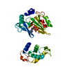
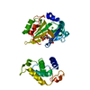
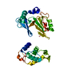
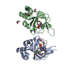
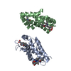
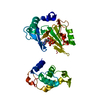
| Title | Crystal structure of myelin protein zero-like protein 1 (MPZL1) | ||||||
 Components Components | Myelin protein zero-like protein 1 | ||||||
 Keywords Keywords | MEMBRANE PROTEIN / receptor / glycoprotein / transmembrane / immunoglobulin | ||||||
| Function / homology |  Function and homology informationcell surface receptor protein tyrosine kinase signaling pathway / cell-cell signaling / focal adhesion / structural molecule activity / cell surface / plasma membrane Function and homology informationcell surface receptor protein tyrosine kinase signaling pathway / cell-cell signaling / focal adhesion / structural molecule activity / cell surface / plasma membraneSimilarity search - Function | ||||||
| Biological species |  Homo sapiens (human) Homo sapiens (human) | ||||||
| Method | X-RAY DIFFRACTION / SYNCHROTRON / MOLECULAR REPLACEMENT / Resolution: 2.746 Å | ||||||
 Authors Authors | Yu, T. | ||||||
 Citation Citation | Journal: Biochem. Biophys. Res. Commun. / Year: 2018 Title: Structural and biochemical studies of the extracellular domain of Myelin protein zero-like protein 1 Authors: Yu, T. / Liang, L. / Zhao, X. / Yin, Y. | ||||||
| History |
|
- Structure visualization
| Structure viewer | Molecule: MolmilJmol/JSmol |
|---|
- Downloads & links
-Download
| PDBx/mmCIF format | 6igo.cif.gz | 134.9 KB | Display | PDBx/mmCIF format |
|---|---|---|---|---|
| PDB format | pdb6igo.ent.gz | 105.6 KB | Display | PDB format |
| PDBx/mmJSON format | 6igo.json.gz | Tree view | PDBx/mmJSON format | |
| Others |  Other downloads Other downloads |
-Validation report
| Arichive directory | https://data.pdbj.org/pub/pdb/validation_reports/ig/6igoftp://data.pdbj.org/pub/pdb/validation_reports/ig/6igo | HTTPS FTP |
|---|
-Related structure data
| Related structure data |  6igtC 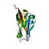 6igwC  3oaiS S: Starting model for refinement C: citing same article ( |
|---|---|
| Similar structure data |


-Links
 PDBj
PDBj


- Assembly
| Deposited unit | 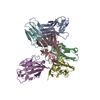
| ||||||||
|---|---|---|---|---|---|---|---|---|---|
| 1 | 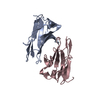
| ||||||||
| 2 | 
| ||||||||
| 3 | 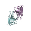
| ||||||||
| Unit cell |
|
-Components
| #1: Antibody | / Protein zero-related Mass: 15220.012 Da / Num. of mol.: 6 / Fragment: UNP residues 36-162 Source method: isolated from a genetically manipulated source Source: (gene. exp.) Homo sapiens (human) / Gene: MPZL1 / Production host:  Escherichia coli BL21(DE3) (bacteria) / Strain (production host): BL21(DE3) / References: UniProt: O95297 Escherichia coli BL21(DE3) (bacteria) / Strain (production host): BL21(DE3) / References: UniProt: O95297 |
|---|
-Experimental details
-Experiment
| Experiment | Method: X-RAY DIFFRACTION / Number of used crystals: 1 |
|---|
- Sample preparation
| Crystal | Density Matthews: 2.47 Å3/Da / Density % sol: 50.16 % |
|---|---|
| Crystal grow | Temperature: 293 K / Method: vapor diffusion / Details: 100mM Tris-HCl pH 8.5, 1.15M (NH4)2HPO4 |
-Data collection
| Diffraction | Mean temperature: 100 K / Serial crystal experiment: N |
|---|---|
| Diffraction source | Source: SYNCHROTRON / Site: SSRF  / Beamline: BL19U1 / Wavelength: 0.9778 Å / Beamline: BL19U1 / Wavelength: 0.9778 Å |
| Detector | Type: DECTRIS PILATUS 6M / Detector: PIXEL / Date: May 14, 2017 |
| Radiation | Protocol: SINGLE WAVELENGTH / Monochromatic (M) / Laue (L): M / Scattering type: x-ray |
| Radiation wavelength | Wavelength: 0.9778 Å / Relative weight: 1 |
| Reflection | Resolution: 2.746→50 Å / Num. obs: 22192 / % possible obs: 99.7 % / Redundancy: 3.4 % / Rpim(I) all: 0.056 / Net I/σ(I): 11.87 |
| Reflection shell | Resolution: 2.746→2.85 Å / Num. unique obs: 1700 / Rpim(I) all: 0.373 |
- Processing
| Software |
| ||||||||||||||||||||||||||||||||||||||||||||||||||||||||
|---|---|---|---|---|---|---|---|---|---|---|---|---|---|---|---|---|---|---|---|---|---|---|---|---|---|---|---|---|---|---|---|---|---|---|---|---|---|---|---|---|---|---|---|---|---|---|---|---|---|---|---|---|---|---|---|---|---|
| Refinement | Method to determine structure: MOLECULAR REPLACEMENT Starting model: 3oai Resolution: 2.746→48.291 Å / SU ML: 0.39 / Cross valid method: FREE R-VALUE / σ(F): 1.97 / Phase error: 28.71
| ||||||||||||||||||||||||||||||||||||||||||||||||||||||||
| Solvent computation | Shrinkage radii: 0.9 Å / VDW probe radii: 1.11 Å | ||||||||||||||||||||||||||||||||||||||||||||||||||||||||
| Refinement step | Cycle: LAST / Resolution: 2.746→48.291 Å
| ||||||||||||||||||||||||||||||||||||||||||||||||||||||||
| Refine LS restraints |
| ||||||||||||||||||||||||||||||||||||||||||||||||||||||||
| LS refinement shell |
|
